Defect reconstruction of the flank represents a complex surgical challenge due to the paucity of suitable recipient vessels and the limited availability of local soft tissue, particularly in patients with low body mass index.
The following preoperative considerations were considered:
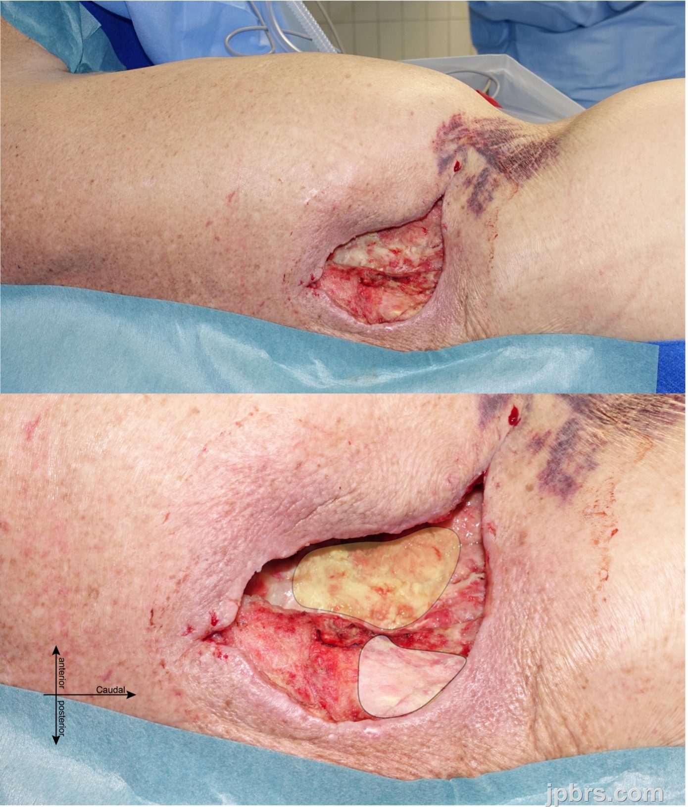
1. Defect characteristics: A full-thickness soft tissue defect measuring 12 × 6 × 5 cm on the right flank resulted from chronic osteomyelitis of the ribs following nephrectomy. The defect involved exposed ribs and peritoneum, necessitating robust vascularized tissue coverage.
2. Reconstructive requirements: The surgical objective was to achieve stable coverage using well-perfused tissue with sufficient volume to obliterate dead space, protect exposed structures, and resist infection recurrence.
3. Evaluation of flap options: Given the defect depth and anatomical location, several reconstructive options were evaluated:
3.1. Local flaps: Local or regional rotational flaps were deemed unsuitable due to the defect’s proximity to the spinal axis, which limited tissue mobility, the patient’s minimal subcutaneous fat reserves, and the defect's complex three-dimensional configuration.
3.2. Tissue expansion: Tissue expansion was ruled out due to the presence of active infection (osteomyelitis and peritoneal contamination), the need for prompt reconstruction, and the associated risk of infection-related complications with delayed techniques.
3.3. Pedicled Transverse Rectus Abdominis Muscle (TRAM) Flap: A pedicled TRAM flap was considered but excluded due to unfavorable anatomical conditions, including a measured distance of 32 cm between the pedicle origin and the superior border of the defect, which exceeded the flap’s reliable arc of rotation.
3.4. Free Anterolateral Thigh (ALT) Flap: A free ALT flap, potentially incorporating part of the vastus lateralis muscle, was evaluated for its volume potential. However, the absence of suitable recipient vessels in the immediate vicinity of the defect would have necessitated interposition grafting for both the artery and the vein. Moreover, the defect’s location cranial to the level of the iliac recipient vessels would have required retrograde arterial flow. These limitations made the free ALT flap an unsuitable option for reconstruction in this case.
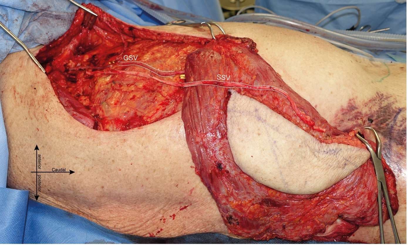
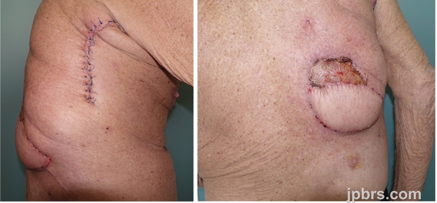
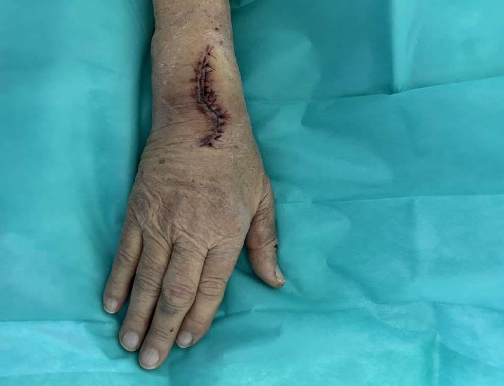
4. Selection of Ipsilateral Myocutaneous Latissimus Dorsi Sliding Flap: Preoperative planning ultimately favored a myocutaneous LD sliding flap from the ipsilateral side. This flap provided sufficient volume, a robust type V vascular supply via the thoracodorsal vessels [1], and the option for pedicle extension through interposed autologous vein grafts to reach the cranial aspects of the defect. The latissimus dorsi muscle’s anatomical proximity, broad surface area, and pliability made it particularly suitable for conforming to the irregular three-dimensional geometry of the flank. Furthermore, pedicle extension allowed for vascular reach while maintaining physiologic anterograde arterial flow.
The decision-making process involved extensive discussions with the patient, focusing on the advantages and disadvantages of each reconstructive option, anticipated outcomes, potential complications, and the impact on quality of life. Additionally, the patient was informed about the potential for intraoperative adjustments to the planned procedure should a more suitable option arise.