如何引用本文
Paula Ejdier, Dogu Aydin. Case 6 (2024): Revolving Door Flap. Journal of Plastic, Breast & Reconstructive Surgery. 2024.
开放获取。 © 2024 作者。由Journal of Plastic, Breast & Reconstructive Surgery发表。本病例版权由作者与期刊共同持有;作者可将自己的病例用于教育、演讲及社交媒体。
继续阅读
Journal of Plastic, Breast & Reconstructive Surgery·经同行评审的开放获取外科病例报告同行评审 · 开放获取

Paula Ejdier, Dogu Aydin. Case 6 (2024): Revolving Door Flap. Journal of Plastic, Breast & Reconstructive Surgery. 2024.
开放获取。 © 2024 作者。由Journal of Plastic, Breast & Reconstructive Surgery发表。本病例版权由作者与期刊共同持有;作者可将自己的病例用于教育、演讲及社交媒体。
继续阅读
Paula Ejdier1, Dogu Aydin1
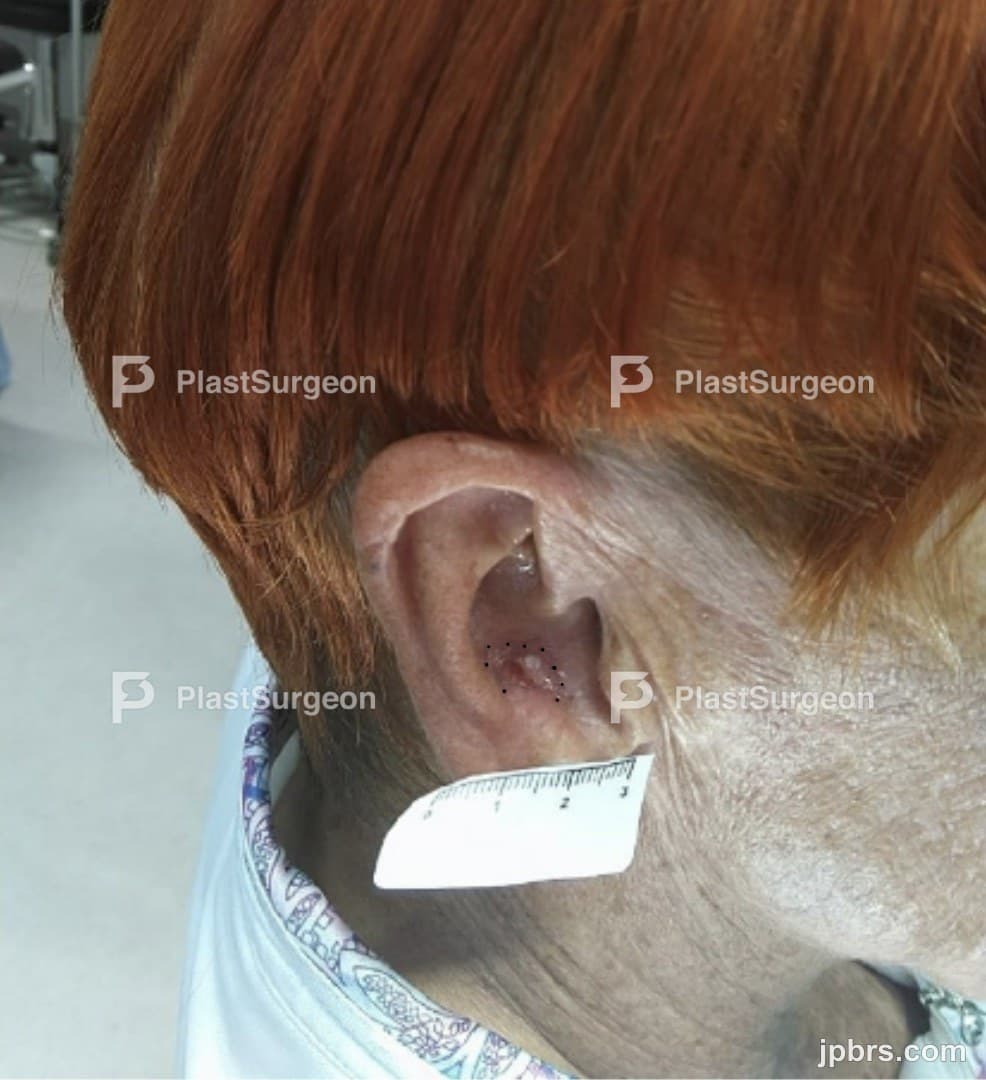
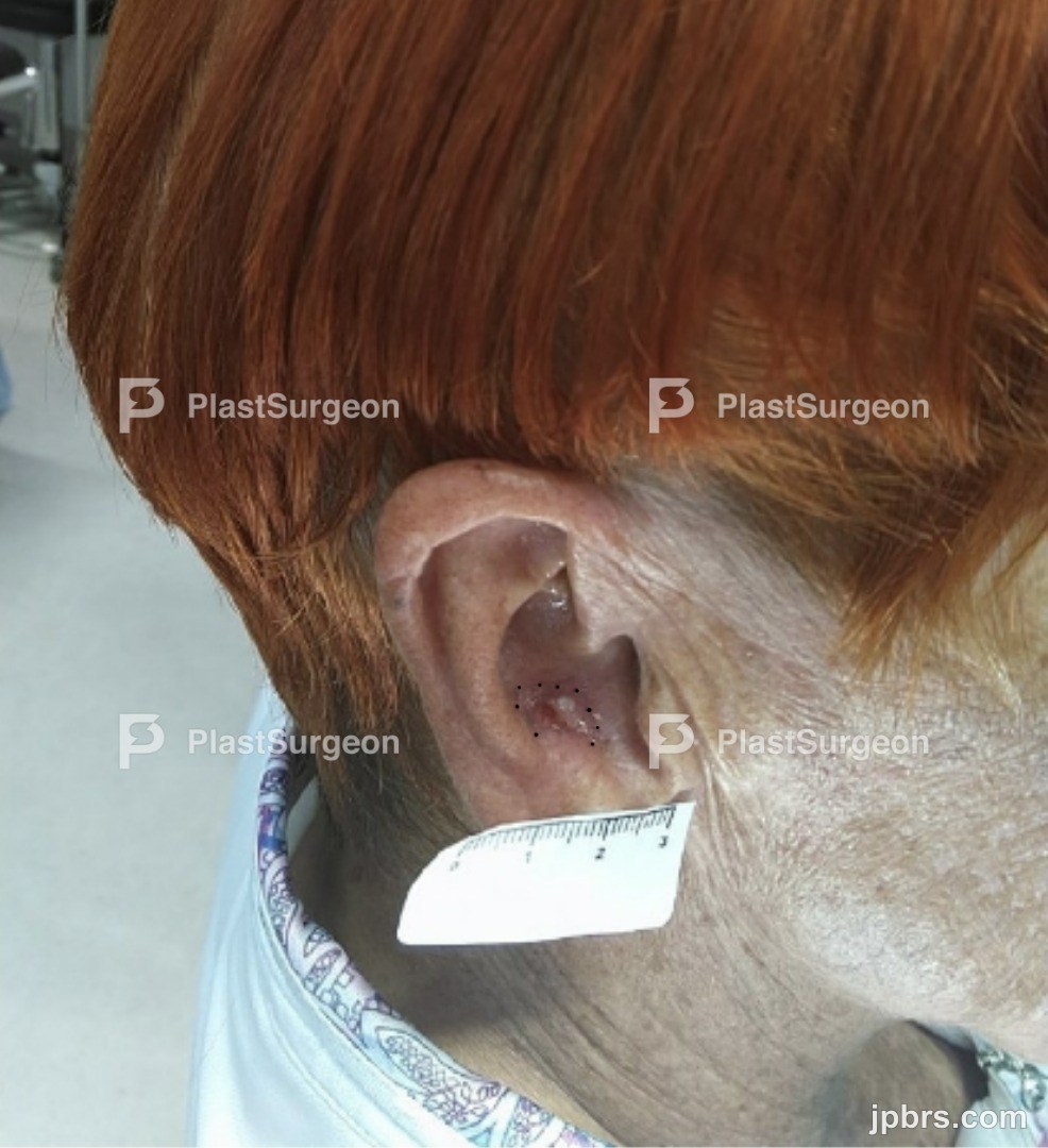
78 year old female referred to Plastic Surgery Department by local ENT-doctor after punch biopsy findings of Squamous cell carcinoma in situ, with questionable stromal invasion on right ear concha. Tumor debut 1 month prior with since fast growth.

 BeforeAfter
78 year old female referred to Plastic Surgery Department by local ENT-doctor after punch biopsy findings of Squamous cell carcinoma in situ, with questionable stromal invasion on right ear concha. Tumor debut 1 month prior with since fast growth.
The patient presents a physical and cognitive well preserved condition. Skin: Clearly visible ulcerated tumor on right ear concha of approx.12x8mm and a central cartilage island of approx.4mm with erosion. Adherent and not displaceable from the underlying tissue. The skin on the back of the ear is intact and without signs of ingrowth. Overall right ear is seen slightly red and swollen (compared to left ear). Very tender when palpated. Lymph nodes: no pathologic lymph nodes were palpable in the region
Squamous cell carcinomas (SCC) present a more aggressive growth pattern compared to Basal cell carcinomas, with a potentially higher risk of metastasis. The tumor of the patient presents a clinical suspicion, as well as in the light of the fast growth and questionable stroma invasion on punch biopsy, of being a SCC rather than SCC in situ. The patient is informed about treatment options in the form of surgery or radiotherapy, as well as the associated advantages, disadvantages and possible complications respectively related. The patient preferres surgery. The patient is planned for an Excision with a 5 mm margin incl. cartilage + perioperative freeze histopathology in order to determine a radical margin before a reconstruction of the defect. Due to the area of surgery, two possible reconstruction methods are considered 1] Revolving Door flap (RD flap) or also known as Postauricular Island flap. 2] Full skin graft, harvested from the e.g preauricular, clavicular or upper arm region. Given the well preserved condition of the aesthetically aware female patient, and in light of an autoimmune precondition, the decision falls upon the local RD flap. Reducing areas of scars, and leaving potential infection risk to only one site.
1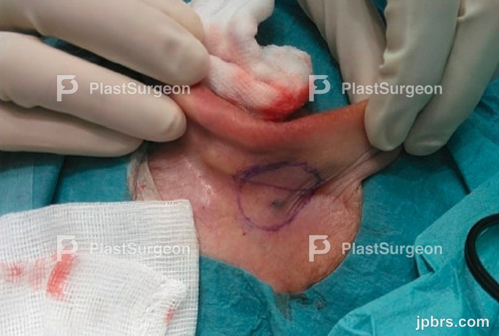
Excision area is outlined with a 5mm safety margin. Under local anesthetic (Lidocaine + Adrenalin) para-auricular and locally, the tumor is excised incl. underlying cartilage. Freeze sections are harvested from margin boarders 12, 3, 6, 9 o’clock + the base and send for perioperative rapid histopathology. Analysis show tumor cells 9 o’clock – why further 5mm margin locally in that section of the defect is excised. Thereafter no signs of malignancy are found corresponding to all the margin boarders + base. Reconstruction part can therefore begin.
2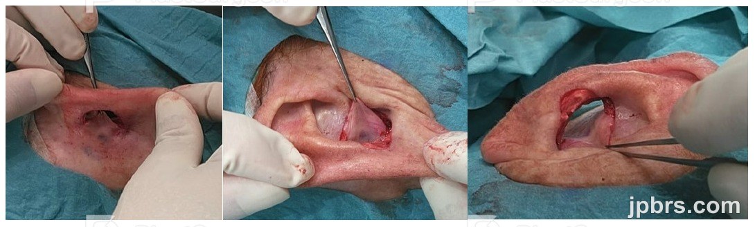
The RD flap is drawn posteriorly with; the ½ circle defined by the remaining retro-auricular skin according to the defect and second ½ circle duplicating the other in size. The midline is defined by the postauricular sulcus.
3
1.stage: The flap is raised taking into account the blood supply stalked throughout the midline of the flap The medial dermal half is pulled forward, and undergoes a rotational movement (like a revolving door) that changes its position from “postauricular” to “antero-auricular”. Now covering the antero-medial half of the frontal defect.
4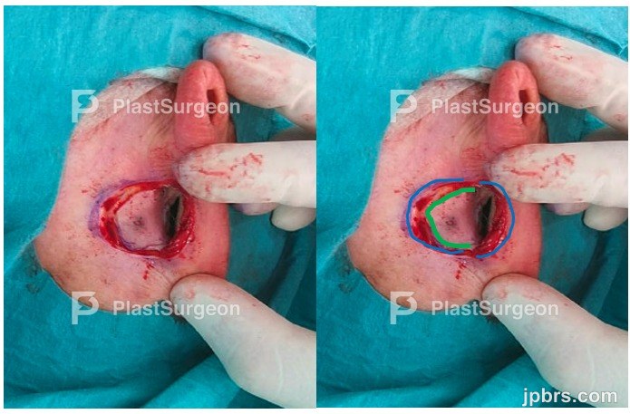
The antero-medial part of the flap is attached superficially using a continuous-suture, Prolene 5-0
5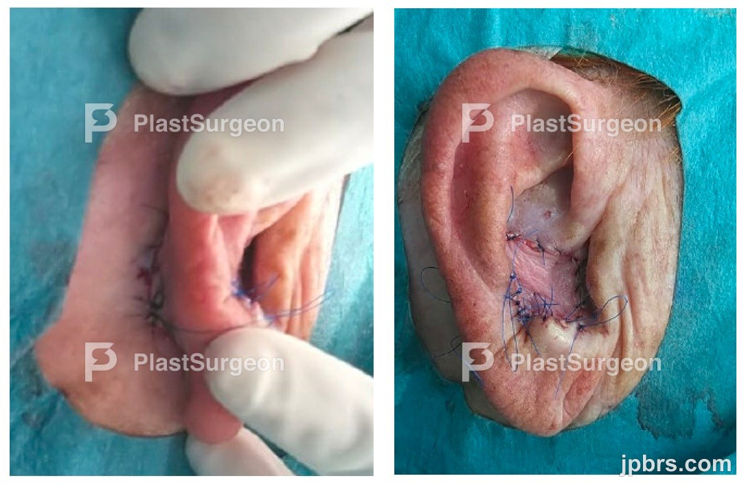
2.stage: Postauricular lateral dermal half is undermined, only at the edges, and pulled forward through the defect (green). Now in a frontal position, it is then attached to the antihelix. The remaining edges are aligned together posteriorly (blue) recreating a new sulcus line.
6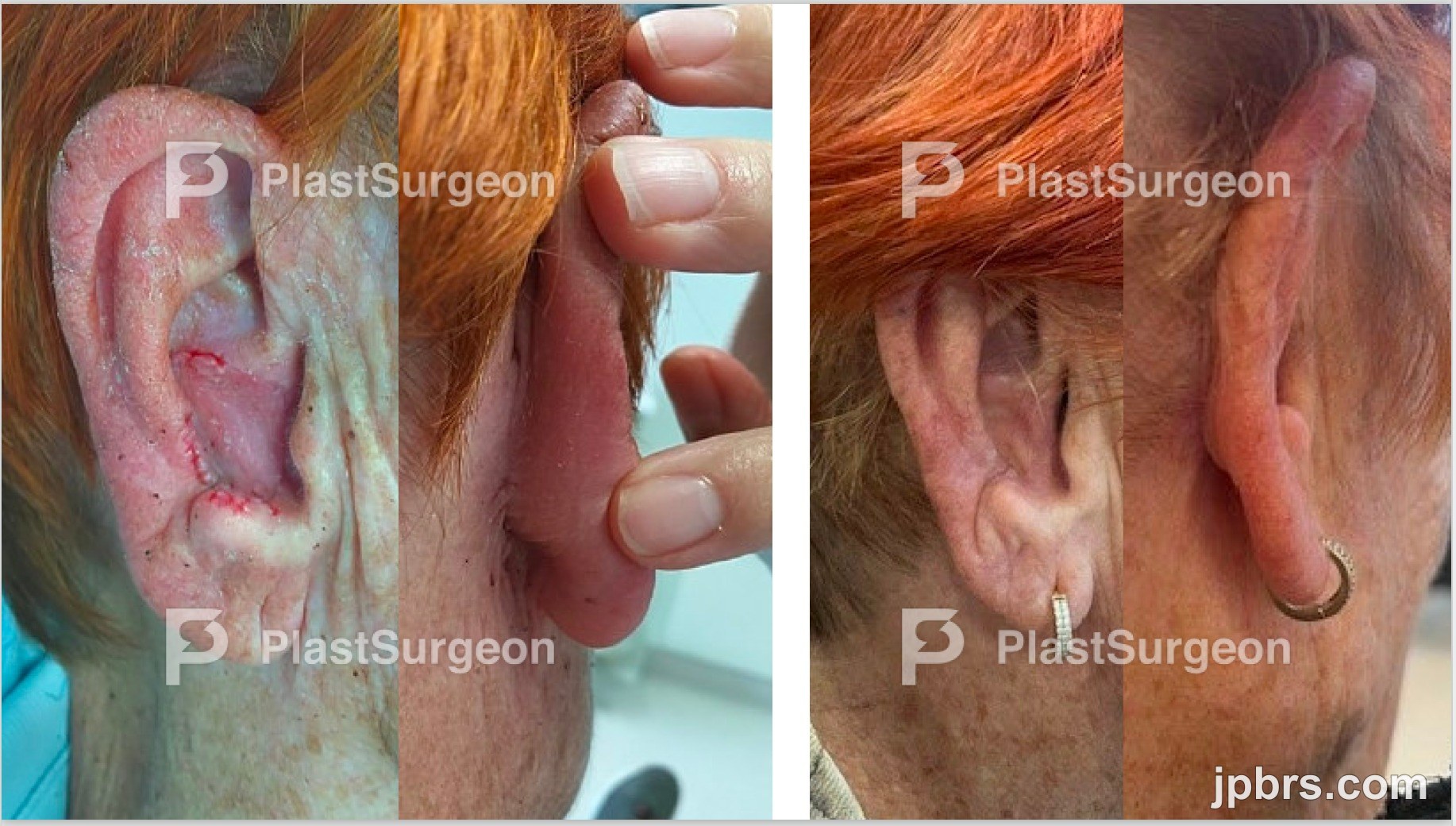
The donorsite posteriorly (left) is closed superficially using a continuous-suture, Prolene 5-0. The antero-lateral part of the flap (right) is attached to antihelix superficially using a continuous-suture, Prolene 5-0. Few supporting single stitches are made here-and-there. Wound dressing: Posteriorly is applied MicroporeM3 on the scar. Anteriorly is applied a sponge to create a compression to the flap and attached by MicroporeM3 (NB no sutures are used to fixate the wound dressing)
7Wound check 2 weeks after surgery (left). The wound dressings are removed. No signs of irritation, infection nor necrosis. Patient is satisfied. The flap had at no point shown signs of complications. Follow up 3 months after surgery (right). The patient is fully satisfied with the cosmetic result with no reports of functional nor other discomforts.

Histopathology showed a radically removed SCC infiltrating the perichondrium deeply but not entering the cartilage itself, with no further vascular nor neural ingrowth. The patient is invited for a final clinical 1year follow up and will thereafter be dismissed from our department with a successful outcome.