如何引用本文
Case 8 (2023): Removal of an axillary lymphnode. Journal of Plastic, Breast & Reconstructive Surgery. 2023.
开放获取。 © 2023 作者。由Journal of Plastic, Breast & Reconstructive Surgery发表。本病例版权由作者与期刊共同持有;作者可将自己的病例用于教育、演讲及社交媒体。
继续阅读
Journal of Plastic, Breast & Reconstructive Surgery·经同行评审的开放获取外科病例报告同行评审 · 开放获取

Case 8 (2023): Removal of an axillary lymphnode. Journal of Plastic, Breast & Reconstructive Surgery. 2023.
开放获取。 © 2023 作者。由Journal of Plastic, Breast & Reconstructive Surgery发表。本病例版权由作者与期刊共同持有;作者可将自己的病例用于教育、演讲及社交媒体。
继续阅读
A 65-year-old male with a palpable lump in his right axilla. Admitted from the department of hematology to perform a lymphadenectomy diagnose for lymphoma. The patient had lost 6 kilos of weight through the last 3 months and his health was beginning to deteriorate.
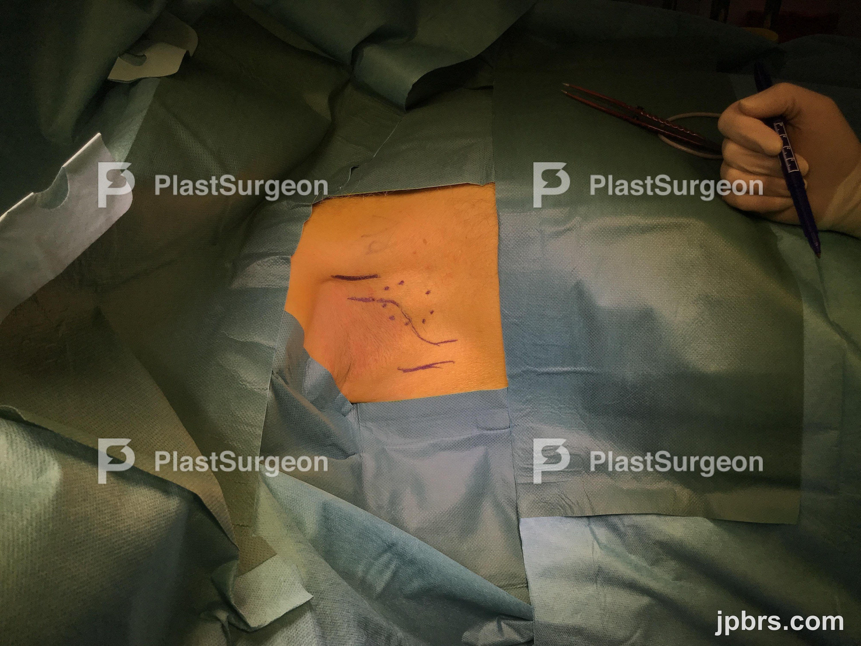
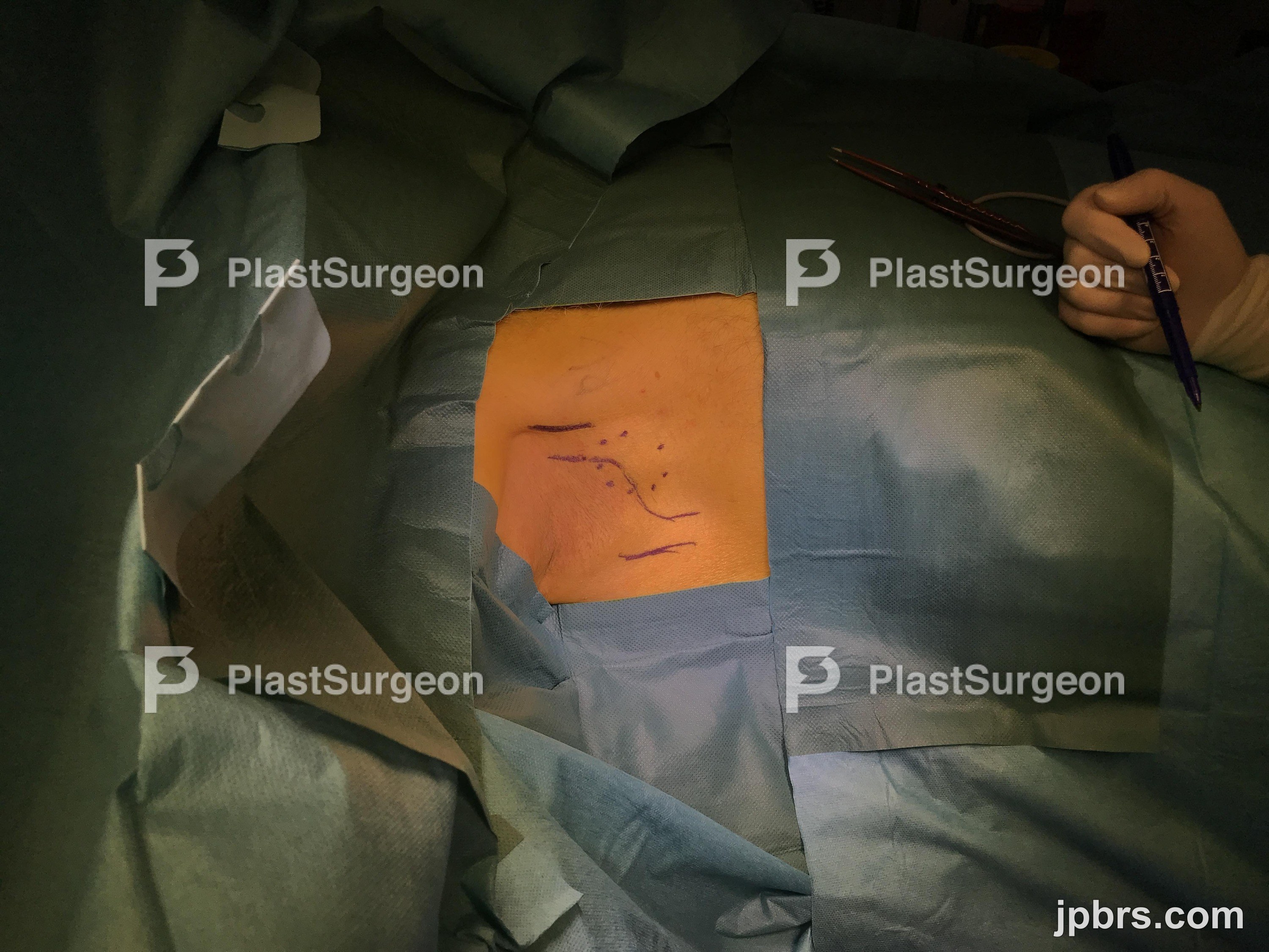 BeforeAfter
A 65-year-old male with a palpable lump in his right axilla. Admitted from the department of hematology to perform a lymphadenectomy diagnose for lymphoma. The patient had lost 6 kilos of weight through the last 3 months and his health was beginning to deteriorate.
When removing a lymphnode in the axilla, one has to consider that the scar tissue may develop into a contracture. Therefore a lazy-s incision should be performed from the pectoral muscle, going diagonal just below the hair area, and advance near the latisimus dorsi muscle. In case there is to be performed a later radical lymphnode dissection, one should remove the incision in 1-2mm margins and use the same lasy-s incision (often elongating the incision to reach all of the lymphnodes). Another important aspect is to send the tissue just in saline solution (NaCl solution), as the pathologist need to assess for lymphoma using microscopy. The lymph node may not be sent in formalin (formaldehyde), as this solution damage the tissue for assessment.
1
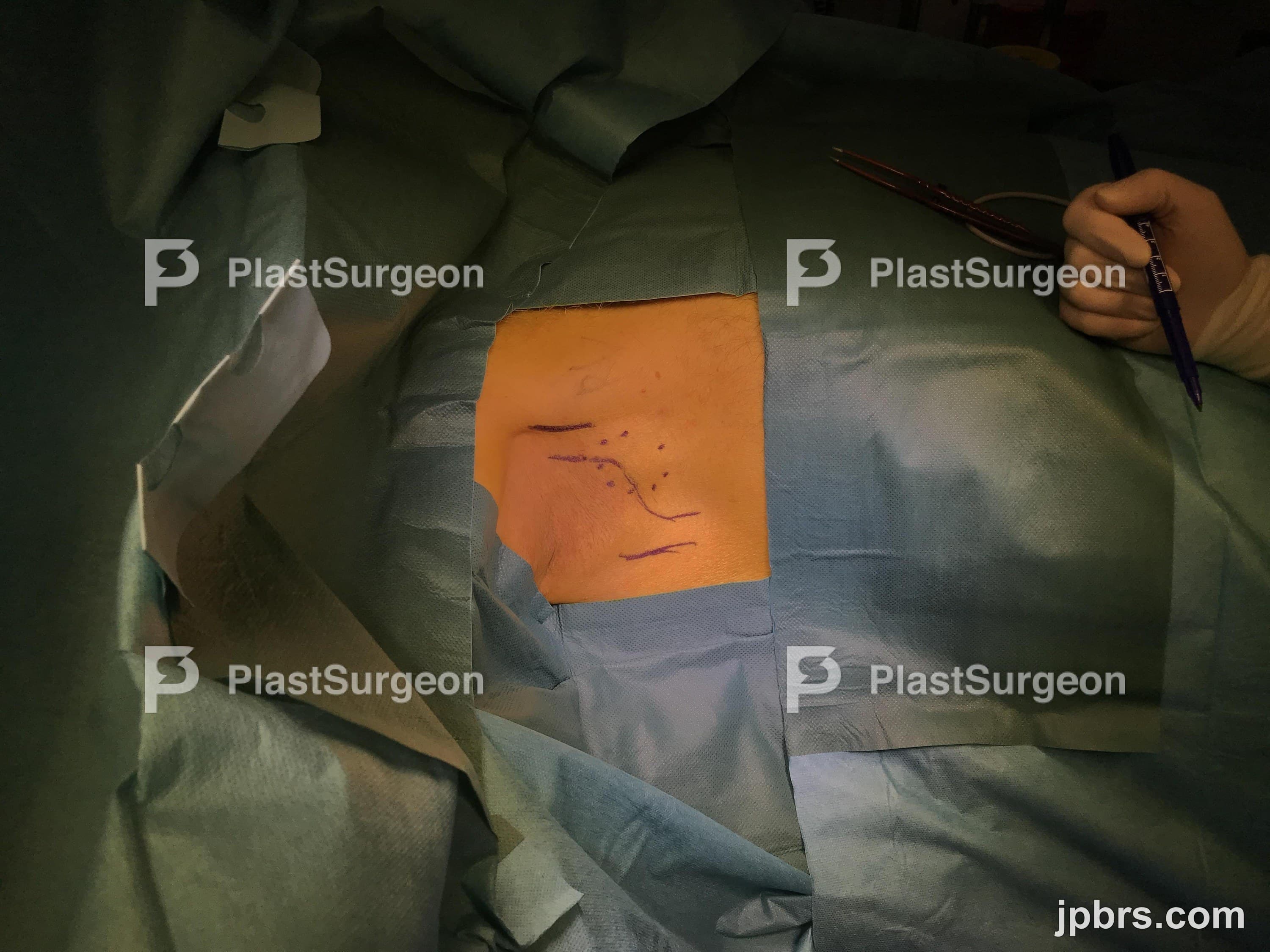
The pectoral muscle is marked anteriorly. The latissimus muscle is marked posteriorly. The palpable lymphnode is marked centrally A lazy-S incision is drawn reaching the near-center of the
2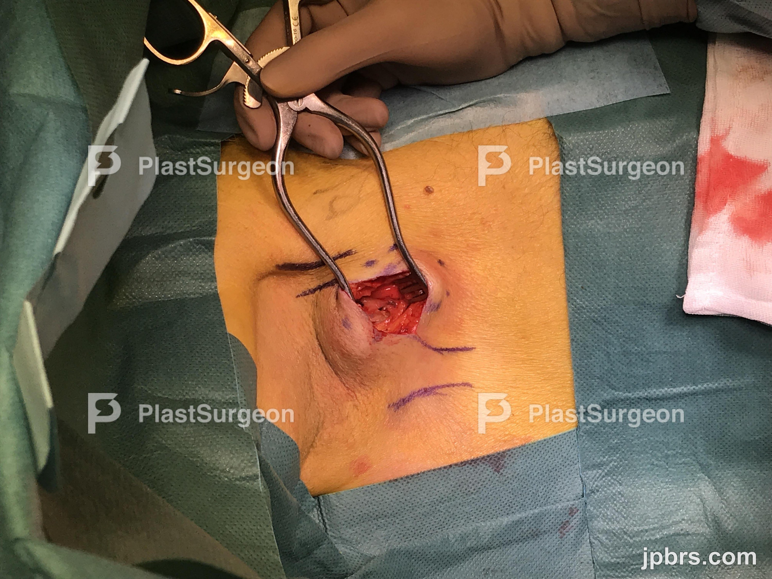
Incision through skin and subdermal tissue
3
When reaching the axillary fat, self-holding retractors may be helpfull to view of the axillary content.
4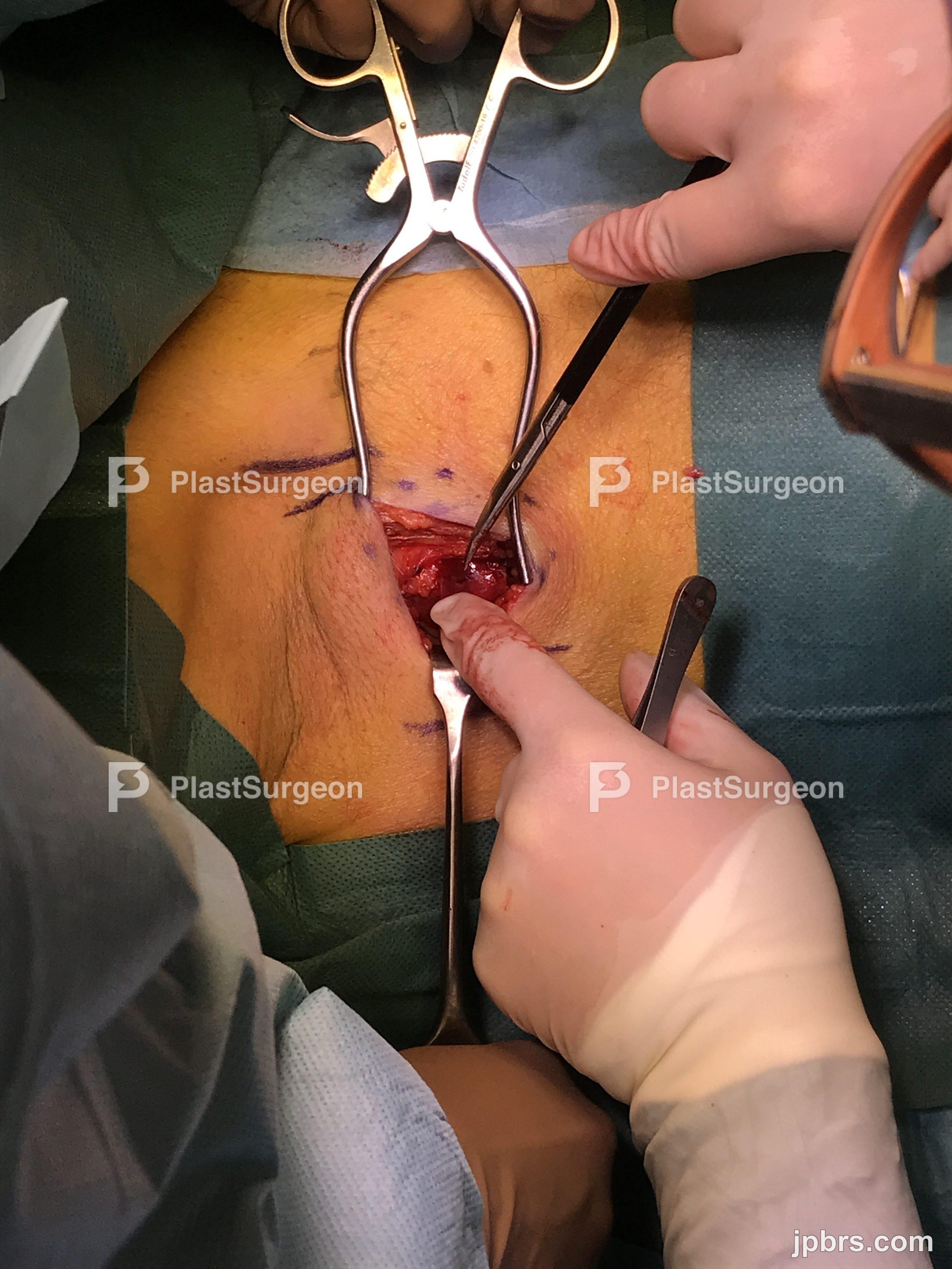
Small bleeding vessels are closed using bipolar diathermia. The lymphnode is dissected very close to the capsule using the bipolar diathermia, thereby ensuring no harm to nerves or larger vessels.
5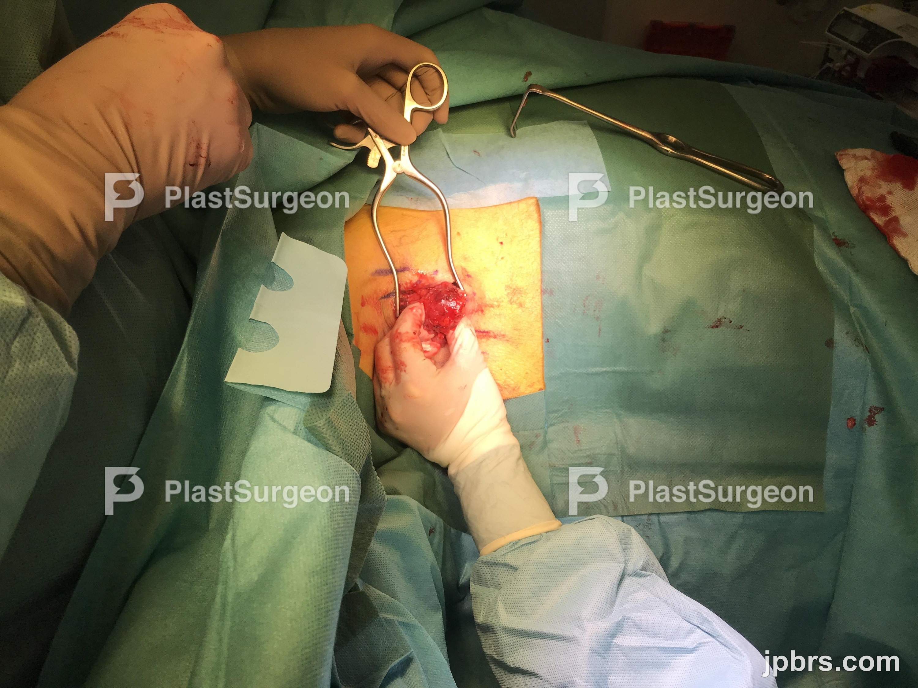
The Lymphnode is removed using a combination of metzenbaum scissors and bipolar diathermia. Keep the dissection close to the capsule of the lymphnode. Use retractors to obtain a better view. Use forceps to handle the lymphnode, holding in the capsule, without damaging it.
6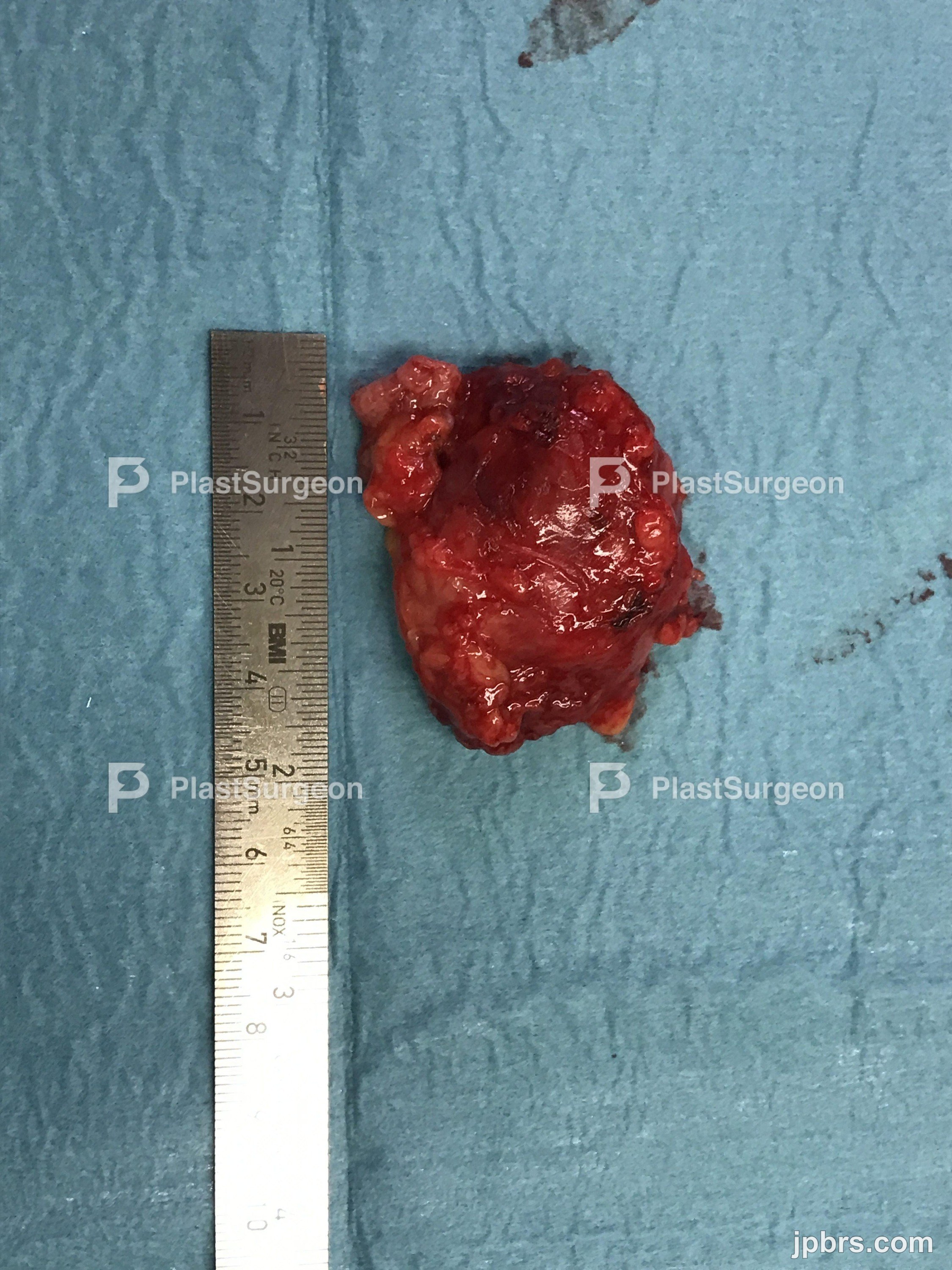
The lymphnode is succesfully removed. The capsule is intact, this ensures no lymphoma cells or cancer cells left behind in the operative field.
7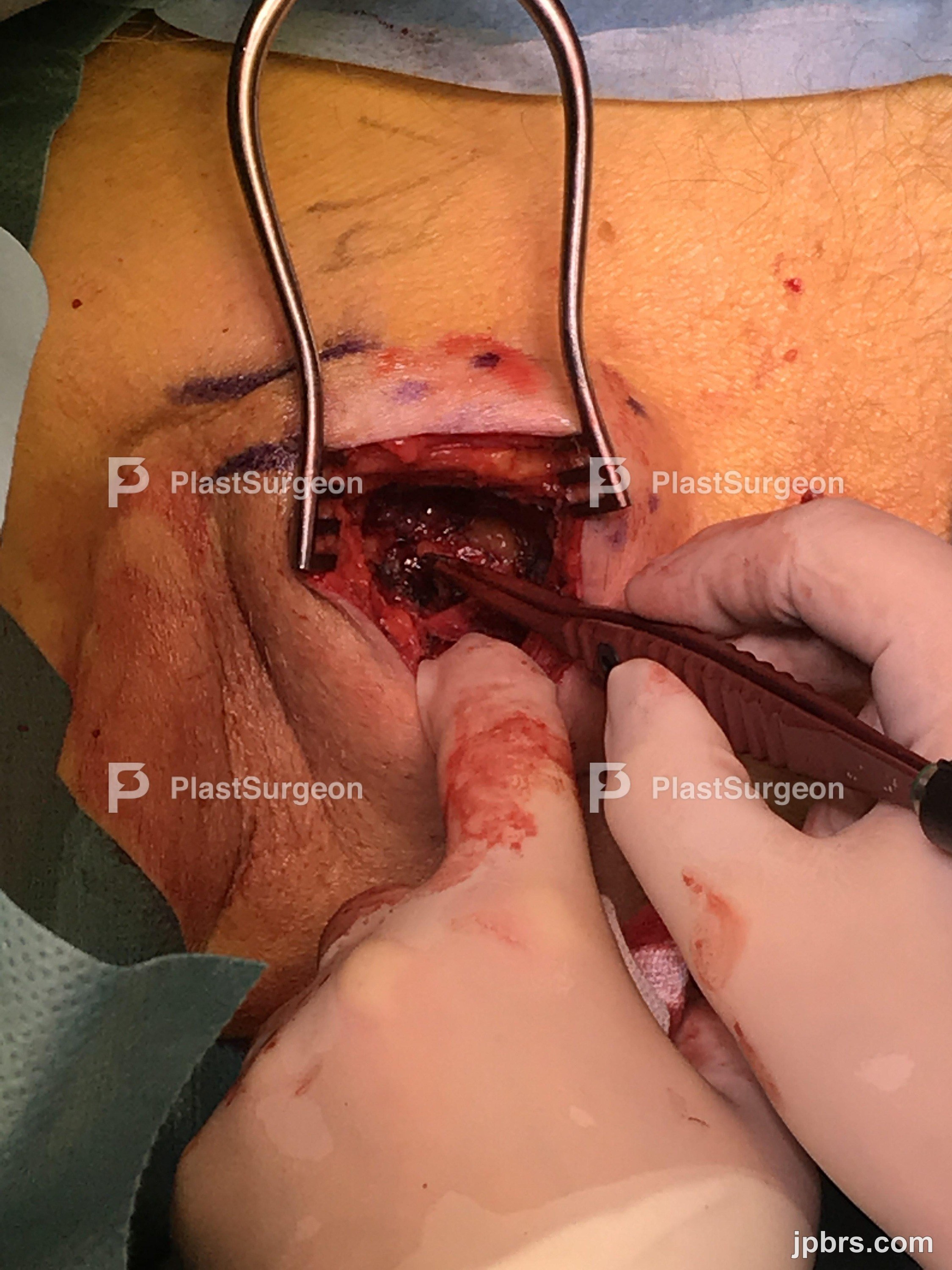
The lymphnode measures 4x3x3 cm in size. The capsule is intact. Some minor fatty tissue is left in the capsule.
8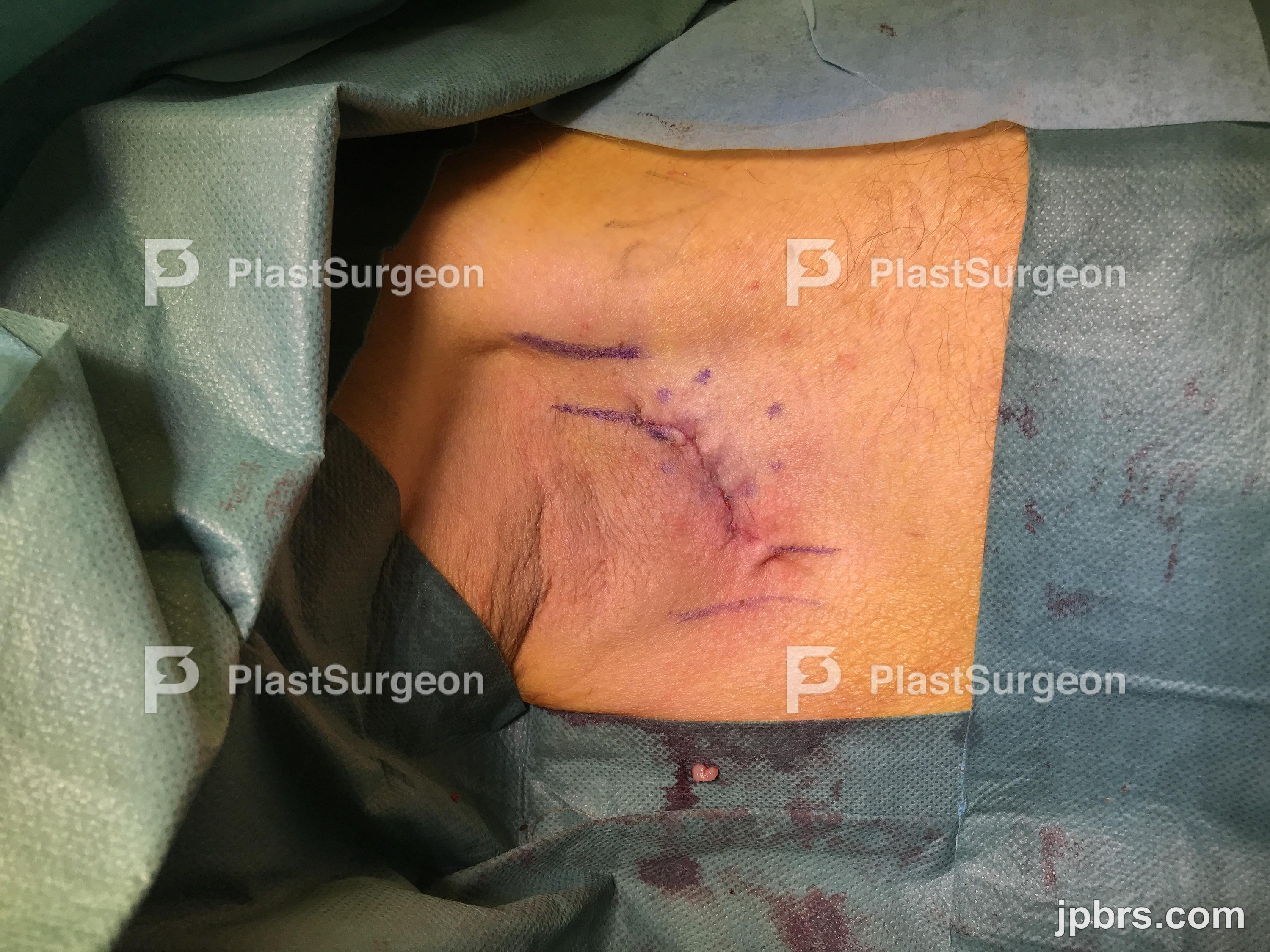
Gently and thoroughly hemostasis is always important
9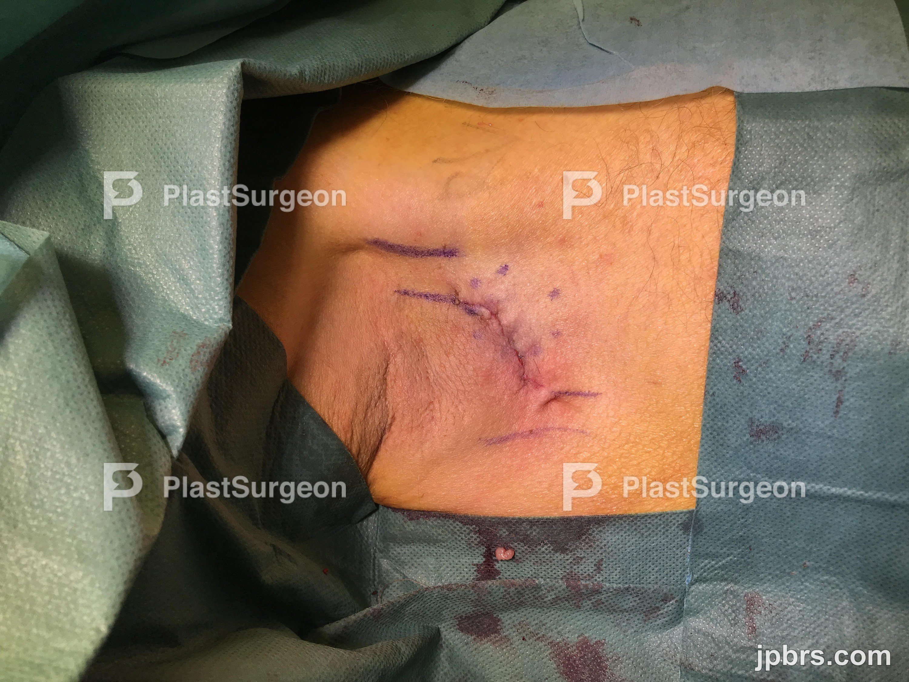
The patient had the skin further closed/secured using micropore plaster. Compression was applied to the axilla using gauze and mefix plaster. The patient was discharged later the same day. No active sports to be performed for 2 weeks.