Reconstructive surgery faces a significant challenge when addressing complex and extensive soft tissue loss in the lower extremity. Use of combined free flaps offers a promising strategy for one-stage reconstruction of such defects. The following preoperative considerations were taken into account:
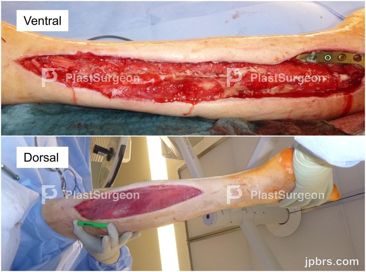
1. A soft-tissue defect measuring 35x4cm was present on the ventral side of the lower left leg. In the proximal section of the defect, there was exposed plate osteosynthesis and tibia. In the distal portion of the defect, the functional structures such as tendons and bones were not exposed.
2. Considering the configuration of the soft-tissue defect and the patient’s high functional demand due to dependency on orthotic support (peroneal splint), the utilization of a split-thickness skin graft in the distal portion of the ventral defect was rendered possible but inadequate to fulfil the functional requirements. Consequently, the decision was made to proceed with full-thickness tissue replacement using a free autologous flap.
3. Based on the potential need for future bone revision procedures, such as removal of the osteosynthesis plates and screws, use of a fasciocutaneous flap was preferred over a muscle flap (e.g., latissimus dorsi free flap) for defect reconstruction.
4. Upon assessment of all available options, and taking into account the patient’s slim nutritional status, we considered that a viable option would have been to use an extended anterolateral thigh (ALT) free flap reaching up to 35 cm in length. However, preoperative perforator mapping using Duplex ultrasonography identified insufficient distal ALT perforators.
5. Another option identified during pre-operative planning was the use of a bilateral ALT-flap up to 35 cm in length. At this stage a decision was made to intraoperatively check for distal ALT-perforators and change to bilateral ALT-flap if perforators were found to be insufficient.
During surgery, a third, and more promising option, arose. Although there were insufficient distal ALT perforators, a significant anteromedial thigh (AMT) perforator was available. To circumvent the aforementioned perfusion challenge associated with the unilateral ALT flap, we ultimately opted to perform a combined ALT and AMT free flap, allowing for primary closure of the donor-site. This option not only allowed complete defect reconstruction, but also simultaneously guaranteed adequate perfusion, thus, reducing donor-site morbidity. For the more superficial dorsal defect, split-thickness skin grafting was performed.
The patient was provided with detailed information regarding the surgical procedure, which included a clear explanation of the potential risks associated with free flap surgery. These risks included the possibility of partial or total flap loss, as well as complications at the donor-site, such as seroma, hematoma, dehiscence, and loss of strength. The importance of close monitoring of the flap during the initial post-operative to quickly detect and address any potential complications was also explained. Specifically, according to our hospital’s protocol, flaps are monitored hourly during the first 48 hours after the surgery. Furthermore, the possibility of modification of the initially planned procedure intra-operatively if a better option becomes available was also explained.
1