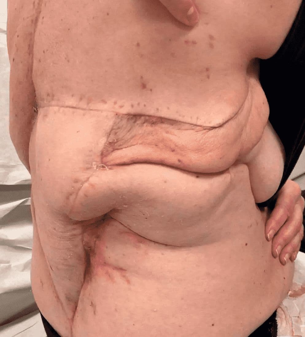
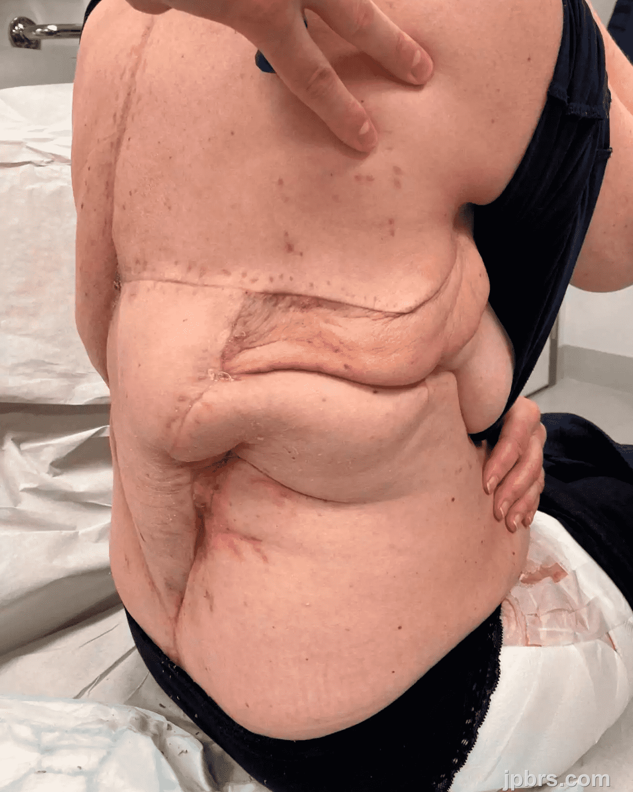
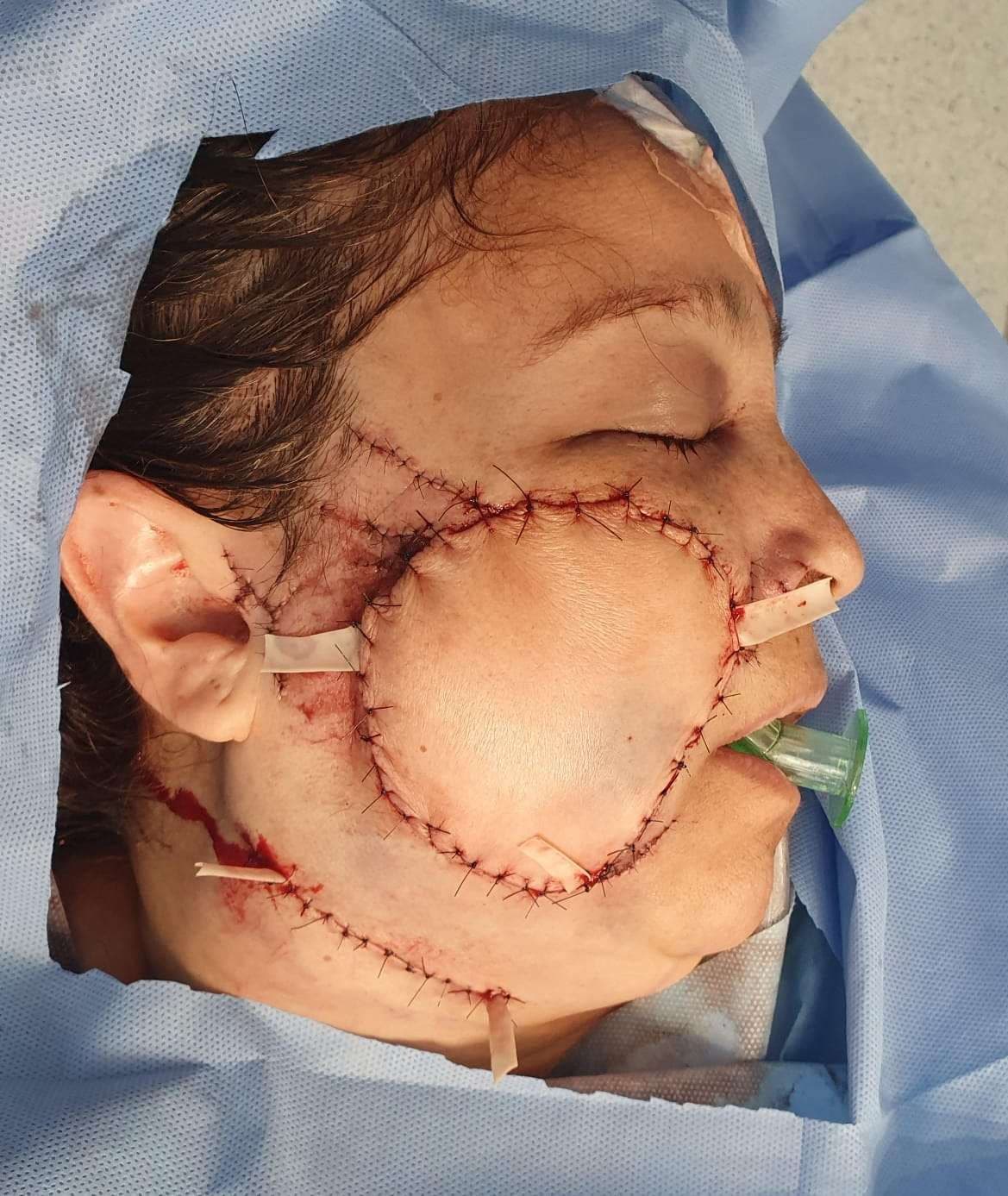
The patient is previously healthy, has no past medical history. She developed a bulge on the back at T12 level. MRI and biopsy showed chondrosarkoma. She was operated at Karolinska University Hospital in May 2022 with sarkoma resection, spinal fixation T 10 to L3, primary skin closure, and post- operative radiotherapy.
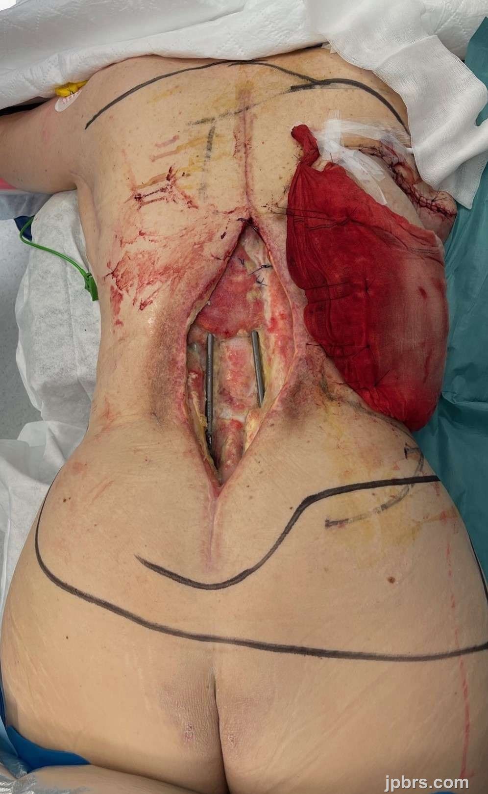
After 2 years the patient presented with osteoporotic reabsorption of bone, fusion/collapse of L1-L2, and rupture of a shaft of the previous fixation. The patient underwent again surgery by the spine surgeon at our hospital in July 2024 with extraction of the previously done fixation, and a new longer fixation was performed T5 to L5. Beacuse the soft tissues ware previously irradiated, a left pedicled muscle latissimus flap was performed to have better coverage of the upper potion of the defect. The wound ruptured and got infected, with expsure of the spinal fixation material; the patient received targeted IV antibiotics and the wound which was 22x10 cm was covered temporarily with a VAC.
In October 2024 at our hospital, we decided to achieve coverage of the wound and re-do the spinal fixation. There were no valid recipient vessels for a free flap near the defect, and no local flaps were possible because the tissue around the wound was very damaged by radiotherapy.
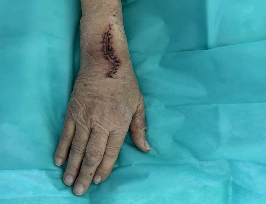
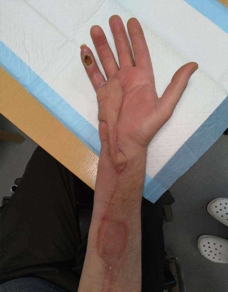
Description of the surgery: We raised av left sided free radial flap (21+7 cm), at the same time as a big right sided ALT (34x15 cm) including vastus lateralis was raised, and the right thoracodorsal vessels in the axilla were exposed. All the superifical veins of the forearm were thrombosed including the cephalic vein. Thus, a 20 cm graft of saphenous vein was harvested from the right thigh.
The Radial flap was hooked up to the axilla, radial artery to thoracodorsal artery, larger Vena Comitans to the serratus branch vein. The saphenous vein graft was anastomosed to the thoracodorsal vein with a vein ring.
The ALT flap was detached from the thigh, flushed with heparin, and kept in iced water. The patient was turned from supine position to side position, with the right side up. In the meantime the radial flap got congested. The ALT was anastomosed to the distal side of the radial artery, and the ALT vein to the saphenous vein graft. The congestion of the radial flap improved (presumably because of flow through) and the ALT was very well blood supplied. Two doppler cooke probes were placed distal to each arterial anastomosis.
The ALT was wrapped in a moist gauze and stabilized to the skin nearby the defect. avoiding tension to the pedicle. The patient was now turned into prone position, and the spine surgeons extracted the infected/exposed fixation material and performed again spinal fixation. The ALT was inset now in the defect.
The patient was immobilized on an air madress in side position for 1 week. Both flaps healed with no complications. The ALT donor site was reoperated several weeks after because of split transplant graft loss. The patient now can walk.