Keywords: Squamous cell carcinoma, adipofascial, turnover flap, finger, defect, reconstruction
Authors: Lukas Kure-Rosenberg, MD Magnus Balslev Avnstorp, MD Nicco Krezdorn, MD, Chief. Institution: Department of Plastic Surgery, Zealand University Hospital, Denmark
Abstract
Reconstruction of finger defects presents unique challenges, particularly in those with complex anatomical involvement. We present a case involving an immunosuppressed patient with a larger late diagnosed SCC, the excision of- and subsequent reconstruction- utilizing an adipofascial turnover flap (APTF), that offers a reliable and versatile option for addressing these challenges, while preserving hand function and aesthetics. A 74-year-old male with a history of kidney transplantation and long-standing immunosuppressive therapy presented with a highly differentiated SCC on the dorsum of the right fourth finger. The lesion, measuring 40 × 35 mm, extended to the proximal and distal interphalangeal joints. Surgical management included two stages: tumor excision with clear margins, followed by secondary reconstruction utilizing an APTF combined with a split-thickness skin graft (STSG). Preoperative and perioperative Doppler assessments guided flap design and ensured adequate vascularity. The APTF provided robust soft-tissue coverage of the exposed extensor tendon, with adequate perfusion confirmed by Doppler. The STSG demonstrated partial take but effectively covered the underlying APTF. Postoperative assessments were followed 30, 48, and 106 days postoperatively. Range of motion (ROM) showed postoperatively some limitations in the affected finger, particularly in the PIP and DIP joints, but functionality was aimed preserved with ongoing therapy. We demonstrate in this clinical case, how APTF is an effective and versatile reconstructive option for complex dorsal finger defects. Its robust vascular supply, adaptability, and minimal donor site morbidity make it particularly advantageous in immunosuppressed patients such as the presented. This case highlights the importance of careful planning, perioperative adaptability, and postoperative rehabilitation to optimize outcomes.
Patient medical history
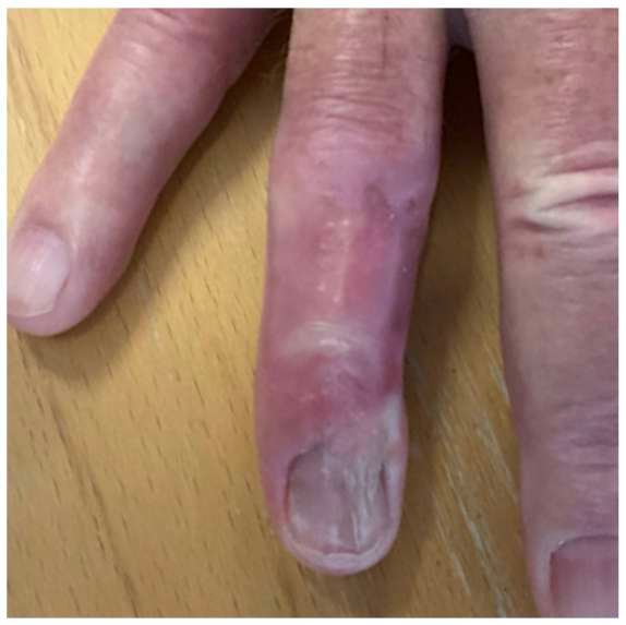
A 74-year-old male was referred to the Department of Plastic-Surgery, Roskilde, Zealand University Hospital, Denmark, fall 2024 by private plastic surgery for treatment of a lesion of the right hands fourth finger. The patient had a history of kidney transplantation and was on immunosuppressive therapy, followed regularly by the dermatology department for skin checkups. The patient had reported a persistent lesion on the dorsum of the right fourth finger for the last 4–5 years. Previously, clinically interpreted as a Verruca Vulgaris and had been recommended over-the-counter treatments without improvement. Medical records indicated cryotherapy treatments since 2018, which were also ineffective. Following establishment of squamous cell carcinoma (SCC) diagnosis via biopsy, it was deeming necessary for hospital-based treatment.


Before and After
Patient examination
During initial evaluation, a poorly differentiated SCC with a multilobulated appearance and some central ulceration, measuring 40 × 35 mm was identified on the dorsum of the right fourth finger. The tumor extended to and were involved with the skin of the proximal interphalangeal joint (PIP) and distal interphalangeal joint (DIP), with encroachment toward the volar side, involving both ulnar and radial aspects. The patient demonstrated normal sensation and capillary refill distal to the tumor and retained full finger mobility. He described the pain as sharp and intermittent. Due to the tumor’s size, adherence to deeper structures was challenging to assess pre-operatively but was preliminarily considered non-adherent to the tendons.
Pre-operative considerations
Given the extent of the defect and its proximity to critical structures, several factors were evaluated to ensure optimal functional and aesthetic outcomes. Alternative reconstruction options, including a metacarpal arterial perforator flap (mAPF), were considered. However, a two-stage adipofascial turnover flap (APTF) was chosen for the following reasons:
Vascularity and Versatility:
The APTF relies on the adjacent capillary network and subdermal plexus for vascularization, ensuring consistent perfusion without the need for extensive dissection or reliance on a single perforator. This minimizes the risk of vascular compromise. Additionally, the flap can be rotated, folded, or inverted to effectively cover deep defects with exposed structures, such as tendons or bone, providing excellent versatility in reconstruction. (Ref: 1)
Tendon Preservation:
By utilizing local tissue with minimal donor site morbidity, the APTF provides effective biomechanical support to exposed peritendinous structures. It ensures adequate soft-tissue coverage while preserving range of motion and mobility without adding bulk or compromising adjacent anatomical structures. (Ref: 2)
Technical Simplicity:
Compared to the metacarpal arterial perforator flap, the APFT requires less extensive dissection and preserves the vascular anatomy of the hand, which may be critical in future surgical interventions. While the mAPF remains a valuable option for dorsal hand and finger reconstructions due to its robust vascularity and pliable tissue, the APFT was preferred in this case because it reduced donor site morbidity—a significant advantage in an immunosuppressed patient. (Ref: 3)
Perioperative Adaptability:
Perioperative assessments also considered the possibility of transitioning to amputation if the lesion involved deeper structures, including the peritendon or bone. Intraoperative examination revealed no macroscopic involvement of these critical structures, allowing pathology to ensure radical margins on HE section and the reconstruction to proceed as initially planned thereafter. The decision to avoid amputation preserved the patient’s digit, which was crucial for maintaining functional outcomes.
Conclusion:
The APTF was chosen as the most appropriate reconstructive option, balancing surgical efficacy, patient safety, and long-term functionality. The ability to adapt the surgical plan perioperatively, including consideration of amputation if warranted, ensured that the approach remained responsive to intraoperative findings. (Ref: 4)

Surgical Preparations and drawing
A digital nerve block was administered using 5 mL of local anesthesia (LA) without adrenaline, targeting all nerve supplies to the right fourth finger, just distal to the metacarpophalangeal joint (MTP). The surgical site was cleansed with an antiseptic solution to achieve sterile conditions. A sterile finger tourniquet was fashioned from a cut surgical glove to maintain hemostasis during the procedure.

1st Stage Procedure: Excision of tumor
Timeout and starting from the ulnar side, incision and dissection were performed with careful visualization and preservation of nerves and vessels, proceeding to the extensor peritendon, which was exposed after tumor elevation (Figure, left). The peritendon was found to be intact, with no evidence of macroscopic tumor infiltration (Figure, center). Dissection continued within this plane toward the radial side, extending across the proximal interphalangeal (PIP) and distal interphalangeal (DIP) joint capsules. On the radial side, the dorsal digital nerve and artery were assessed adherent to the tumor and resected with the tumor specimen, which was orientation-marked and submitted for expedited hematoxylin and eosin (H&E) pathology evaluation. The macroscopically clear defect (Figure, right) was dressed in Jelonet, gauze, and a finger bandage. Follow-up and control were planned accordingly.

Wound Care
The patient was evaluated for wound care in the outpatient clinic 5 days after the first stage surgery. No signs of infection were observed, and the patient reported minimal pain, which was managed effectively with over-the-counter analgesics. Sensory deficits in radial, dorsal, the distal part of the finger was noticed as expected.

2nd Stage Procedure: Reconstruction utilizing an Adipofascial Turnover Flap
Eleven days after tumor excision, radical tumor removal was confirmed microscopically. Twenty days post-excision, secondary stage with closure of the defect, now measuring 40 × 40 mm, was performed.
During operative preparation, dressing removal and cleansing of the defect revealed degeneration and weakening of the extensor tendon in the distal part of the defect, resulting in partial mallet finger deformity. This was addressed during the procedure. Preoperative Doppler examination identified signal from the dorsal aspect of a. digitalis palmaris propria proximal to the defect, including signal dorsally, confirming adequate perforator flow for reconstruction.
Under sterile conditions and local anesthesia as previously, but without tourniquet, the procedure was performed following time-out. The extensor tendon was repaired using a 4-0 Prolene Kessler suture technique, restoring the tendon to its normal position.
An incision was made over the radial-dorsal aspect of the skin proximal to the defect, extending distally over the dorsum of the metacarpophalangeal joint (MTP). The skin flap was elevated with respect for the underlying structures on an ulnar base (Figure, left).
Perioperative Doppler confirmed perforator flow from the dorsal segment of the a. digitalis palmaris propria at A7, with the strongest signal originating from the radial side perforator. The adipofascial layer was mobilized from the ulnar side toward the proximal part and radially, with regards to preserving the perforator. The flap was divided longitudinally to ensure complete coverage of the tendon across the defect (Figure center) and was fixed distally with 4-0 Vicryl simple inverted sutures.
A 2.5 × 5 cm split-thickness skin graft (STSG) was harvested from the right cubit, with primary closure of the donor site. The graft was manually meshed and secured over the APFT using 4-0 Nylon interrupted sutures, ensuring preservation of the vascular supply to the underlying flap (Figure right). The wound was dressed with Jelonet, foam, a custom splint, and a finger bandage. Postoperatively, the patient was scheduled for bolus dressing removal in the outpatient clinic seven days after surgery.

Post-Surgery follow-ups
The patient was evaluated in the outpatient clinic seven days after the second stage surgery for dressing removal. Moderate to significant swelling of the finger was observed, with normal vascular conditions distal to the surgical site. The skin graft appeared fragile with only partial take (Figure, top left), but provided adequate coverage of the APFT (which itself provided cover to the extensor tendon). In consultation with the patient, a decision was made to proceed with conservative healing. The patient was referred to ergonomical therapy and the reconstruction was subsequently evaluated at 30 days (Figure, top right), 48 days (Figure, bottom left), and 106 days (Figure, bottom right) postoperatively, demonstrating progressive improvement.
Pearls
Vascularity and Perfusion:
The flap relies on a robust capillary network and subdermal plexus, providing consistent vascularization. (Ref: 1)
Flap Design and Versatility:
The ability to rotate, fold, or invert the flap allows for coverage of complex defects with exposed tendons or bones, ensuring functional and aesthetic results. (Ref: 1)
Minimized Donor Site Morbidity:
The flap is harvested from local tissues, which reduces the risk of complications and preserves hand functionality. (Ref: 4)
Perioperative Flexibility:
Maintain readiness to adapt intraoperatively. In this case, transitioning to amputation was not chosen because macroscopic inspection revealed no deeper structure involvement.
Immunosuppressed Patient Considerations:
The flap’s robust perfusion and limited surgical complexity make it an ideal choice for immunosuppressed patients, reducing the risk of infection and delayed healing. (Ref: 1,4)
Pitfalls
Vascular Compromise:
Overzealous dissection or undermining can disrupt the subdermal plexus, leading to flap necrosis. Ensure careful handling of the adipofascial tissue during elevation. (Ref: 1)
Flap Tension:
Avoid excessive tension on the flap during rotation or folding, as this may compromise perfusion and lead to partial necrosis. Proper preoperative planning of flap dimensions is crucial to avoid this issue. (Ref: 3)
Delayed Healing in Immunosuppressed Patients:
Patients on immunosuppressive therapy are at higher risk for wound infections and delayed healing. Proactive infection control and close postoperative monitoring are critical. (Ref: 4)
Post-operative plan
The patient was concurrently referred to and followed by occupational therapy during wound healing follow-up. After achieving satisfactory healing, the patient was discharged from the department of Plastic Surgery and continued regular dermatological follow-up for skin surveillance.
Once healing was complete after around 120 days, a silicone dressing was applied by occupational therapy to prevent hypertrophic scar formation. The patient exhibited persistent sensory deficits in the distal dorsal radial side of the finger but engaged in sensory training using tools such as arthro-rollers, hand trainers, and Dr. Winkler exercises. While the patient was encouraged to perform active, non-weight-bearing blocking exercises, these were not consistently practiced.
Before referral to municipal physiotherapy, the following range of motion (ROM) measurements were documented for the right fourth finger:
The patient’s ROM demonstrated limitations upon discharged. However, ongoing therapy and exercises were recommended to improve functionality further and reduce stiffness.
References
- Cavadas, P. C., Landin, L., & Ibáñez, J. F. (2008). Adipofascial Turnover Flap for Coverage of Dorsal Finger Defects.
- Moojen, D. J., et al. (2007). Treatment of Tendon and Bone Defects in the Hand Using Local Flaps.
- Matsui, Y., et al. (2019). Comparative Outcomes of Perforator Flaps in Hand Reconstruction.
- Sokolich, J. C., & Lin, C. H. (2020). Soft-Tissue Reconstruction of the Hand: Flaps and Techniques.
