Keywords: Nasal reconstruction, perialar flap, crescentic, advancement, PCC in situ.
Authors: Lukas Kure-Rosenberg, MD and Nicco Krezdorn, MD, Chief. Institution: Department of Plastic Surgery, Zealand University Hospital, Denmark.
Abstract
We present the case of a 68-year-old woman with a residual PCC in situ on the nasal dorsum following initial excision and open defect by colleague. The initial pathology revealed incompletely excised changes cranial to the primary defect (12 o’clock position). The patient was booked for re-excision and closure, originally planned with a full-thickness skin graft (FTSG). However, the patient expressed high cosmetic expectations, prompting a change in plan to a local flap-based reconstruction. A perialar crescentic advancement flap was chosen, utilizing the planned re-excision site, which anatomically corresponded to a burrow triangle in the flap design. This case illustrates how integrating oncologic and reconstructive planning can optimize both margin control and aesthetic outcomes.
Patient medical history
A 68-year-old woman was referred for further management of an ulcerating lesion, consistent with biopsy-confirmed squamous cell carcinoma (SCC/PCC) in situ on the nasal dorsum. The patient was routinely seen at dermatological checkups due to being kidney transplanted, and was therefore also recommended surgical management of the lesion. The lesion was initially excised by a colleague and left as an open wound for secondary intention until the final pathological analysis. Pathology demonstrated no invasive tumor, but histopathological evaluation revealed residual SCC in situ/atypical changes at the superior (12 o’clock) margin of the excised area, necessitating re-excision.


Before and After
Patient examination
Upon reexamination, the surgical defect was located centrally on the nasal dorsum, cranial to the supratip area, measuring 12 mm in diameter. The previously excised region had demarcated edges and healthy granulation tissue. No signs of infection or tumor invasion were noted. The re-excision area at the 12 o’clock position was carefully evaluated and integrated into the flap planning.
Pre-operative considerations
Several reconstructive options were considered:
FTSG: Technically simple but suboptimal in cosmetic outcome, especially in this central facial location.
Other local flaps (e.g., bilobed, trilobed): These would not have an advantage in excising residual in situ changes.
Paramedian forehead flap: Overly extensive for this case.
Perialar crescentic advancement flap: This flap addressed the area to be excised in its design, while still providing an optimal balance between coverage, aesthetic blending of scars, and the use of local tissue.
Given the location of the residual tumor area, which matched the position of the superior burrow triangle in the flap design, the crescentic advancement flap was deemed the ideal choice.

Surgical Preparations
Initial lesion (left and central picture) and open wound following primary excision (right picture).
All surgery was carried out in an out-patient setting. The nose was preoperatively numbed with local anesthesia (LA) with adrenaline in a combined nerve block of the infraorbital nerve (V2) and infiltration. The surgical site was cleansed with an antiseptic solution to achieve sterile conditions.

Perioperative planning
A perialar crescentic flap was designed and raised along the alar groove, extending into the melolabial fold. The flap was undermined and advanced medially. The superior burrow triangle, which overlapped with the re-excision site, was performed with a good margin (>3mm) and marked for histopathological analysis. Closure was completed in layers with 5-0 Vicryl and 5-0 Prolene sutures.
Re-excision at the cranial 12 o’clock position contained within the burrow triangle of the chosen flap design (left picture). Excision of the burrow triangle (central left picture). Raising the flap (central right picture), until sufficient tension-free coverage of the widest part of the defect (right picture).

Flap inset and suturing
The reconstruction and flap demonstrated good cosmesis and vascular perfusion immediately postoperatively, with no bleeding or complications. The patient was discharged and scheduled for suture removal at the outpatient clinic 7 days post-surgery.

7-Day Postoperative Follow-up
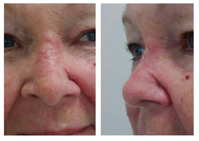
On postoperative day 7, the flap demonstrated excellent perfusion with mild expected edema. Some signs of infection were noted, so the patient started a 5-day oral antibiotic course. Suture removal was carried out at this visit. At the 3-month follow-up, the patient showed minimal scarring with excellent color match and contour. No contractures or distortion were present, and the patient was pleased with the result.

3-Month Postoperative Follow-up
Results 3 months postoperatively, showing complete healing and excellent cosmesis.
Pearls
Accurate Flap Design: Careful planning and design of the perialar crescentic advancement flap, considering anatomical landmarks such as the burrow triangle, ensures optimal tissue alignment and aesthetic outcomes.
Local Anesthesia Efficiency: The combination of nerve blocks and infiltration anesthesia allows for adequate pain control in outpatient settings, facilitating a smoother surgical experience.
Early Detection of Infection: Prompt recognition of early signs of infection and initiation of antibiotic treatment can prevent complications and ensure good flap healing.
Pitfalls
Inadequate Margin Control: Failure to ensure adequate margins during re-excision can lead to incomplete tumor removal, necessitating further interventions and compromising oncologic safety.
Tension on Flap Closure: Excessive tension during flap closure can compromise vascular perfusion, leading to flap necrosis or poor cosmetic results. Adequate undermining and advancement of the flap are crucial to avoid this.
Failure to Consider Patient Expectations: In cases with high cosmetic demands, failing to assess and address patient expectations can result in dissatisfaction, even when the technical outcome is successful.
Post-operative plan
This case demonstrates how re-excision of residual tumor can effectively be combined with aesthetic reconstruction in nasal plastic surgery. The use of a perialar crescentic advancement flap provided a single-stage solution that addressed both oncologic safety and patient satisfaction.
References
- Baker SR. Local Flaps in Facial Reconstruction. 3rd ed. Elsevier; 2014.
- Zitelli JA. The crescentic advancement flap. Arch Dermatol. 1989;125(7):957–959.
- Burget GC, Menick FJ. Aesthetic Reconstruction of the Nose. Mosby; 1994.
- Park SS. Local flaps: cheek and perinasal reconstruction. Facial Plast Surg Clin North Am. 2011;19(1):107–120.
- Rohrich RJ, Muzaffar AR, Janis JE. The Anatomy and Applications of the Nasolabial Flap. Plast Reconstr Surg. 2003;111(2):811–822.
