Case 6: Fingertip reconstruction by a V-Y flap and eponychial flap in a patient with fingertip amputation

Keywords: Fingertip amputation; hand trauma; local flap; eponychial flap; V-Y flap

Authors: Alina Strohmaier, MD, Martina Greminger, MD. Institution: HOCH Cantonal Hospital St. Gallen, Switzerland

Abstract

A 58-year-old male presented with fingertip amputation of the left index finger after a bicycle accident. The injury was classified as Ishikawa Zone 2/Allen Type III, with ulnar oblique pulp defect, subtotal nail bed loss but with intact germinative matrix, and exposed bone. Treatment involved a volar V-Y advancement flap for soft tissue coverage and an eponychial flap to allow visible nail growth. Postoperatively, early mobilization was conducted. At 6-month follow-up, the patient reported good functional outcomes. Examination revealed adequate soft tissue coverage, a normally growing but short nail without a claw-nail deformity, and a stable fingertip. The patient was able to play the piano again. A near-complete range of motion and normal sensation were achieved. This case demonstrates successful fingertip reconstruction using local flaps to restore function and maintain the nail, even when the nail bed is almost completely injured.

Patient medical history

A 58-year-old male patient presented to the emergency department with amputation of the distal phalanx of the second digit on the left dominant hand. The patient sustained the injury while inspecting the brakes of a bicycle, resulting in the second digit impacting the bicycle spoke. The patient is a professional piano player. No regular intake of medication.

Before and After

Emergency presentation of the patient with fingertip amputation (Ishikawa zone 2, Allen Type III) of the left index finger. Initial examination revealed an ulnar oblique amputation of the distal phalanx of the index finger (adominant). The visible part of the nail bed was almost completely injured; however, the germinative matrix remained intact. Sensitivity and blood circulation were preserved. Tendon function was intact for flexors and extensors. Exposed bone was visible. The amputate was available, but was injured in two pieces and therefore unsuitable for replantation. X-rays demonstrated a small radiopalmar dislocated and dorsally malaligned fracture fragment and intra-articular avulsion of the ulnar base.

The fingertip amputation was classified as an Ishikawa Zone 2 or Allen Type III injury (Tos et al. 2024).
In this case, there was an ulnar oblique defect of the pulp, subtotal laceration of the visible nail bed, and a bony defect with bone exposure. With the remaining function of the DIP joint, sufficient attachment of the deep flexor and extensor tendons was assumed.

There are several options to treat the injury, which should be adapted to the patient’s wishes and requirements and taken into account when considering individual treatment. The main goals of treatment are restoration of sensation and durability in the tip and assuring proper bone support to allow nail growth. In addition, an optimized healing time and an acceptable cosmetic appearance are important. Several treatment options for fingertip amputations in general should be considered (Peterson et al. 2014):

1. Replantation of the fingertip: This method can be an effective treatment, especially for sharp Tamai I and II lesions (Venkatramani et al. 2011). In our case, the amputate was split into two parts and was unsuitable for replantation.

2. Secondary wound healing ((semi-)occlusive dressing): Although this treatment method has shown effectiveness for various injury patterns, it yields the most favorable aesthetic and functional outcomes when used for Allen level I injuries (Peterson et al. 2014). One reason for poor aesthetic outcome can be the lack of bony support to the nail. With additional wound contracture this may result in hook nail deformity. Secondary wound healing has its limitations in volar oblique injuries, exposed bone, additional fractures of the distal phalanx, and large soft tissue defects.

3. Bone shortening, eradication of nail matrix, and stump formation with direct wound closure: This method shortens the length of the finger and does not preserve the nail. It is necessary in more proximal amputations, when replantation is not possible. In our case, the stability of the fingertip was crucial; therefore, our goal was to maintain nail growth.

4. Local flap coverage (advancement flaps and neurovascular island flaps): Local flaps are an effective solution for managing fingertip amputations, as they enable maintenance of finger length and provide adequate bone support and soft tissue coverage. These flaps address the palmar defect and should be paired with an eponychial flap to enhance nail length and improve fingertip stability.

1

Wound debridement and planning

Patient in supine position with left arm extended on arm table. Anesthesia with
plexus anesthesia. Sterile disinfection with Octenisept and draping in the usual manner.
Work under loupe magnification (4.5x). Upper arm tourniquet with 250 mmHg.
First, debridement with removal of the bone fragment was performed.
2

VY-flap and eponychial flap

The remaining well-perfused radial skin was successfully mobilized to cover the very distal part of the bone. However, a defect measuring 10×5 mm persisted on the ulnar side of the fingertip. To address this, a V-Y flap was considered to be sufficient and marked on the ulnar side. The skin incision was performed with a scalpel, ensuring careful protection of the vascular-nerve bundle. Circumferential skin transection and flap mobilization were achieved by separating the connective tissue septa from the subcutaneous tissue. By advancing the tissue from the ulnar side, tension-free suturing with Prolene 4-0 was accomplished, resulting in effective soft tissue coverage of the bone.

Key considerations are that the VY-flap is not dissected proximally to the DIP-joint and the neurovascular bundles are not isolated. The flap is mobilized with a Gillies hook by sharply releasing the fibrous septa from the distal phalanx.
3

Suturing and perfusion control

To achieve an aesthetically and functionally pleasing outcome, an eponychium plasty was added to extend the visible part of the nail and enhance its appearance. The incision site and area designated for de-epithelialization were delineated. The available nail pocket measured approximately 4 mm. Deep radial and ulnar skin incisions were made, followed by de-epithelialization of the marked area using a scalpel. Subsequently, the flap was advanced proximally into the de-epithelialized region and secured using Prolene 4-0 sutures.
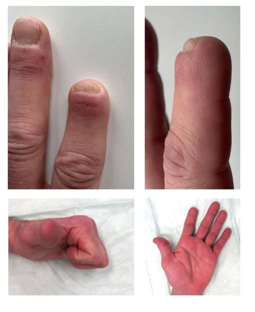
Fingers and hand with redness and swelling
4

Follow up

Follow up with a great result, and a minor size decrease of the finger defect.

Pearls

• An eponychium plasty is worthwhile even when we barely have any visible nail bed remnant from the injury
• The nail is essential for a stable fingertip; in this patient, it was very helpful to play the piano.
• In this instance, the procedure was conducted under regional anesthesia; however, it was feasible to perform the operation using local anesthesia in conjunction with a finger tourniquet.
• Early mobilization and rehabilitation are essential for maintaining range of motion and function.
• Regular follow-up and patient education (hand therapy) are important for monitoring healing and promptly addressing complications.

Pitfalls

• Complications of this operation include nail deformities, loss of pinch strength, loss of or alteration in sensation, cold intolerance, joint stiffness, painful neuromas, cosmetic concerns, and prolonged time away from work.
• Inadequate debridement can lead to infection or poor wound healing.
• Careful flap design and tension-free closure promote optimal healing and minimize complications (flap necrosis).

Post-operative plan

Immediate free mobilization of the finger was advised, accompanied by elevation of the affected limb to mitigate soft tissue swelling. Sutures were removed after 14 days. The patient was regularly seen by an occupational therapist to provide for adequate mobilization and wound healing.
At the clinical follow-up 6 months postoperatively, the patient reported complete pain relief. He was able to play the piano without problems and the fingertip felt stable. During the examination, the scar conditions showed no irritation or swelling. The fingertip showed sufficient soft tissue coverage without axial tenderness. No hook nail deformity occurred. The two-point discrimination at the fingertip was 2-3 mm. aROM MCP 90/0/0° PIP 105/0/0° and DIP 70/0/0°. Force in Jamar handle level 2 left: 40 kg and right: 35 kg.

References

  • Tos P, Crosio A, Adani R. Fingertip injuries and their reconstruction, focusing on nails. Hand Surg Rehabil. 2024;43S. doi:10.1016/J.HANSUR.2024.101675
  • Peterson SL, Peterson EL, Wheatley MJ. Management of fingertip amputations. Journal of Hand Surgery. 2014;39(10):2093-2101. doi:10.1016/j.jhsa.2014.04.025
  • Venkatramani H, Sabapathy S. Fingertip replantation: Technical considerations and outcome analysis of 24 consecutive fingertip replantations. Indian J Plast Surg. 2011;44(2):237-245. doi:10.4103/0970-0358.85345

Contents

Case 6: Fingertip reconstruction by a V-Y flap and eponychial flap in a patient with fingertip amputation