Keywords: Decollement injury, full-thickness skin graft, primary full-thickness skin grafting, lower leg trauma, soft tissue debridement, perforating blood supply, atypical decollement, plastic surgery, trauma management, diabetes, case report
Authors: Laura Emilie Marr Spore, MD., Claes Hannibal Kiilerich, MD. Institution: Department of Orthopedic Surgery, Holbæk Hospital, Denmark
Abstract
An 84-year-old female sustained significant lower leg trauma after a fall on a staircase. Intraoperative evaluation revealed a decollement injury. Defined as separation of the skin and subcutaneous tissue, from the underlying fascia, separating the perforating blood vessels from the subcutaneous tissue—with the affected skin area destined for necrosis, if not acted upon. Prompt debridement and primary full-thickness skin grafting of the affected skin were performed within hours. Early recognition and management are often needed, to avoid devastating injuries. This case highlights a successful treatment, and the need to focus on these injuries, as no clinical guidelines exist.
Patient medical history
The patient, with a history of type 2 diabetes, CKD 5 (Cronic Kidney Disease Stage 5, eGFR < 10, weekly dialysis) and poor skin quality, experienced a fall from standing height on a stone staircase. These factors contributed to a complex decollement injury despite the low-energy trauma. She hit the staircase corresponding to her tibial tuberosity, and a grinding motion during the fall resulted in the visual distal open lesion — only a small proximal wound suggesting where she struck the stairs initially, where the decollement lesion was later discovered to be starting from. The decollement injury is most commenly seen in victims in severe traffic accidents and typically involves opposing, intense forces that tear the skin and the subcutaneous layer away from the underlying fascia, thereby compromising the blood supply to the skin. And is often underdiagnozed and hard to recognize. Decollement lesions are classified into three subtypes: open, closed, and atypical and this case was classified as an open type/atypical type, due to the relatively small open lesion, compared to the extended tissue damage.


Before and After
Patient examination
Preoperatively, the wound was described as a 20 cm inverted Y-shaped lesion located anterolaterally on the distal third of the crus. Underneath the skin flaps, the tibial bone and fascia were visible. No decollement injury was initially suspected, but the 20 cm lesion was later shown to be way smaller than the underlaying 40 cm widespread decollement injury.
Pre-operative considerations
Although no overt decollement or avital tissue was apparent initially, the surgical team maintained the patient’s risk factors in mind. Usually these kinds of injuries are treated at high specialty departments, with a multidisciplinary two-step approach. In this case the patient was booked for a regular wound revision, and the type of injury the propagation of the decollement injury was not apparent before having the patient on the operating table

Mapping out the decollement injury and preparing the full thickness skin graft
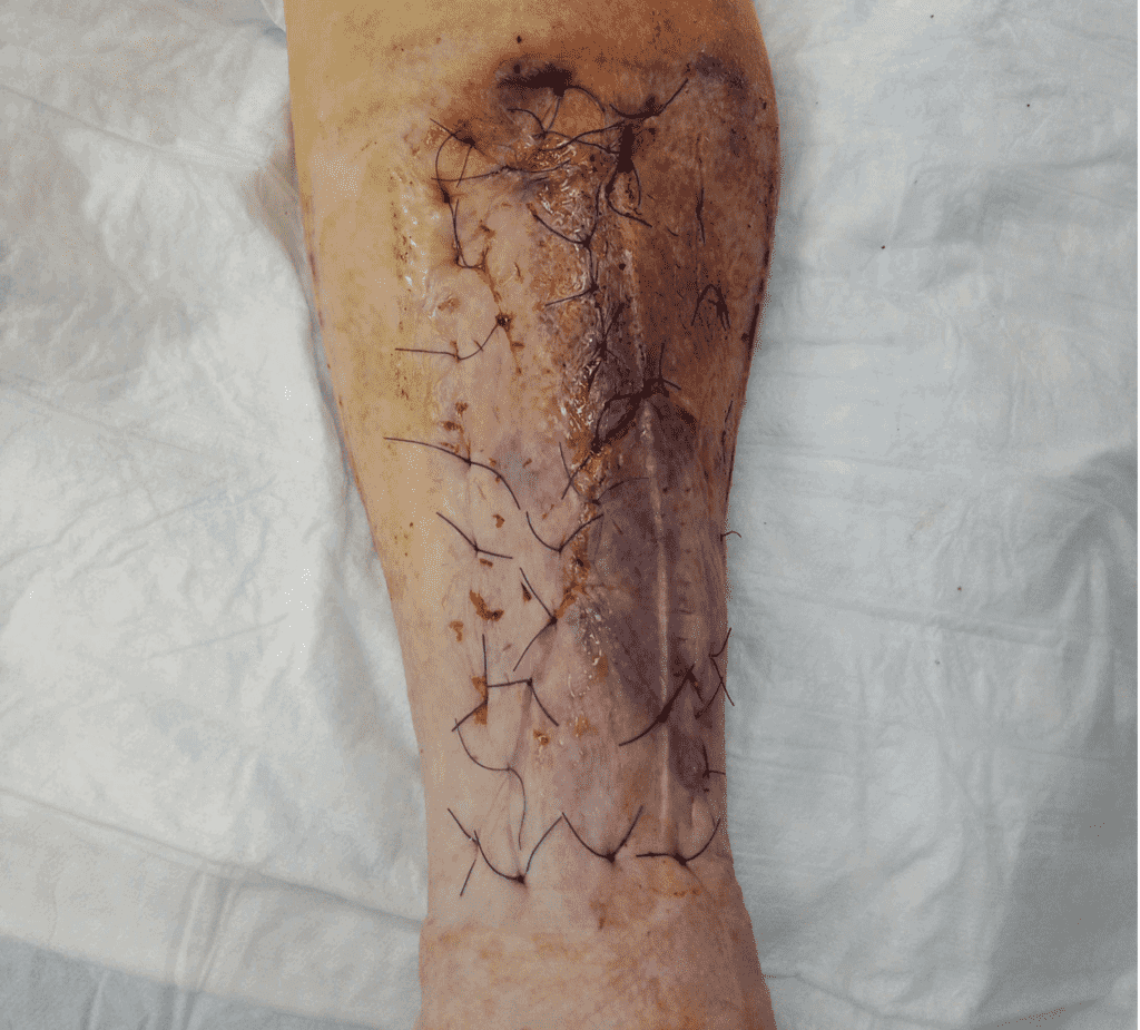
Intraoperative assessment of the injury. An underlying decollement injury was found to be showing a 40 cm propagation of the injury, where fascia and tibial bone are visualized. Avital subcutaneous fat, tissue and debris was mapped out. The wound was thoroughly debrided ensuring a healthy, well-vascularized bed. Meticulous hemostasis was achieved, and wound edges were refreshed as needed. The area was irrigated to minimize contamination.

Preparing and placing the Full Thickness Skin Graft
Primary grafting was performed, using the skin from the traumatized skin flap, and was prepared by removing all of the avital tissue. The skin flap was marked out and made to fit as the donor bed, including both the epidermis and the full dermis. The “recipient” site was marked out

Securing the graft to the donorsite
The graft was accurately aligned and secured onto the recipient bed. With deep sutures that anchored the skin graft to the fascia, around the entire circumference of the graft, so called quilting sutures. And closing the wound shaped as an inverted Y with simple sutures. Small fenestrations were created in the graft to allow for fluid egress to promote adherence and allowing the graft to stretch, covering a larger area. A dressing of “Mepitel one” as the contact layer and several foam dressings as absorbent and compression dressing was stapled onto the skin, to maintain even pressure and immobilize the graft. Lastly a elastic dressing was applied from foot to above the knee, serving to support venous function in the leg of this older lady with nefropathy.

Normal vital tissue
A prototypical example of normal, vital tissue in which, starting from the innermost layer: bone, fascia its perforants, muscle, subcutaneous tissue, fat and cutis.

The mechanism of the decollement injury
The injury occurs when high-impact, opposing forces cause the skin and subcutaneous tissue to separate from the underlying fascia, thereby disrupting the skin’s blood supply. Decollement injuries are typically evident in open skin wounds—but atypical cases may have minimal or no external signs. This subtle presentation can easily mask extensive soft tissue injury, making diagnosis challenging in the initial phase. The illustration showing an open type.

Treatment of the decollement injury
Several treatment strategies favor reusing the skin over the decollement lesion as split- or full-thickness grafts. In the illustrated method, a full-thickness graft with deep quilting sutures promotes revascularization from the fascia. With foam and compression used to minimize hematoma, seroma, and drainage issues. Vacuum therapy has been suggested in the literature as the gold standard.

Follow up
Pearls
Pitfalls
Post-operative plan
References
- Latifi R, El-Hennawy H, El-Menyar A, Peralta R, Asim M, Consunji R, Al-Thani H. The therapeutic challenges of degloving soft-tissue injuries. J Emerg Trauma Shock. 2014 Jul;7(3):228-32. doi: 10.4103/0974-2700.136870. PMID: 25114435; PMCID: PMC4126125.
- Hidalgo DA. Lower extremity avulsion injuries. Clin Plast Surg. 1986 Oct;13(4):701-10. PMID: 3533379.
- Boesen MP, Larsen CF, Elberg JJ. Decollement. Diagnostik og behandling [Decollement. Diagnosis and treatment]. Ugeskr Laeger. 2004 Jan 12;166(3):139-43. Danish. PMID: 14870639.
- Hierner R, Nast-Kolb D, Stoel AM, Lendemans S, Täger G, Waydhas C, Schmitz D, Husain N. Die Décollementverletzung im Bereich der unteren Extremität [Degloving injuries of the lower limb]. Unfallchirurg. 2009 Jan;112(1):55-62; quiz 63. German. doi: 10.1007/s00113-008-1548-z. PMID: 19224101.
- Yuan K, Zhao B, Cooper T, Jin Z, Zhou X, Chen X, Li Z, Gao W, Yan H. The management of degloving injuries of the limb with full thickness skin grafting using vacuum sealing drainage or traditional compression dressing: A comparative cohort study. J Orthop Sci. 2019 Sep;24(5):881-887. doi: 10.1016/j.jos.2019.01.002. Epub 2019 Jan 30. PMID: 30709789.
