Keywords: ala nasi, nasolabial flap, infranasal flap, bullhorn modification, facial reconstruction
Authors: Lukas Kure-Rosenberg, MD, Nicco Krezdorn, MD, Chief. Institution: Department of Plastic & Breast Surgery, Zealand University Hospital, Denmark
Abstract
Reconstructive surgery of the caudal ala nasi poses a particular challenge due to the complex anatomy and high aesthetic significance of the area. The skin in this region is thick, inelastic, and poorly mobile, and the alar contour plays a crucial role in the overall symmetry and three-dimensional shape of the nose. Additionally, local tissue availability is limited, making flap design and execution technically demanding. Two-stage procedures, which are often required in this area, can be disadvantageous as they involve prolonged treatment time, multiple interventions, and temporary aesthetic or functional compromise between stages, potentially affecting patient satisfaction. Preoperative considerations should include a detailed assessment of the defect’s size, depth, and proximity to the alar rim, as well as evaluation of nasal symmetry, skin quality, and patient expectations. It is also important to plan for structural support, such as cartilage grafts, if there is a risk of alar collapse or distortion. This report presents three cases involving patients treated for basal cell carcinoma (BCC) on the caudal part of the nasal ala, following excision with frozen section guidance. Case 1 underwent a traditional two-stage nasolabial flap and demonstrated the subsequent need for revision due to dynamic tension and functional concerns. Case 2 introduces a novel single-stage, medially based infranasal flap – a “modified half bullhorn approach” – tailored to address smaller defects in a single-stage, cosmetically discreet and functionally balanced manner. Case 3 introduces an even larger single-stage, medially based contralateral bullhorn transposition flap with advancement closure – a “modified full bullhorn approach with contralateral transposition” – tailored to address defects involving the floor of the nasal vestibule, also in a single-stage, cosmetically discreet and functionally balanced manner.
Patient medical history
Case 1, a 72-year-old female referred to the Department of Plastic Surgery, Roskilde, Sealand University Hospital, Denmark, in fall 2024, following referral by a dermatologist for surgical treatment of a lesion on the right lower alar edge. Biopsy revealed nodular basal cell carcinoma. The patient had a medical history of type 2 diabetes mellitus, asthma bronchiale, hypertension arterialis essentialis, and was an active smoker. Case 2, a 67-year-old male, was referred by a dermatologist to the Department of Plastic Surgery, Roskilde, in spring 2025 for treatment of a lesion on the lower left alar edge, with biopsy confirming nodular basal cell carcinoma. Case 3, a 82-year-old female, was referred by a dermatologist to the same department in spring 2025 for treatment of a lesion involving the infranasal region with extension into the nasal vestibule and lower left ala. Biopsy revealed nodular basal cell carcinoma. She had a history of type 2 diabetes mellitus.


Before and After
Patient examination
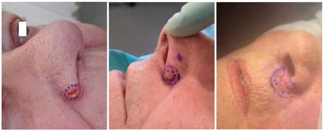
During initial evaluation, Case 1 presented with a lesion with a small central ulceration, measuring 10 × 8 mm. The tumor did not extend to the nasal vestibule. Case 2 presented with an 8 mm lesion, also not involving the nasal vestibule. Case 3 involved a larger tumor measuring 22 × 11 mm. All cases were planned for surgical excision, intraoperative frozen section examination, and reconstruction with a local random flap due to anatomical location.
Case 2: The patient requested minimal intervention. A novel, medially based infranasal flap (modified half bullhorn) was designed. The donor site was closed using nasolabial advancement with a small back-cut to avoid upper lip distortion.
Case 3: Due to patient anxiety and logistical barriers to radiotherapy, a single-stage contralateral transposition flap was planned. Intraoperatively, the defect proved larger than expected. A modified full bullhorn flap with contralateral transposition and additional nasolabial advancement was performed to address the vestibular involvement.
Pre-operative considerations
Case 1: Two-Stage Classical Nasolabial Flap
A classic, minor, two-stage nasolabial flap was chosen by the surgical supervisor. After initial flap inset and pedicle division, with emphasis on recreating the alar edge via a sharp pedicle turn, the patient was seen for 3-month follow-up. Although the cosmetic outcome did not concern the patient, she reported discomfort and functional disturbances during speech and facial motion. The nose was noted to deviate slightly to the contralateral side. A second-stage flap revision was planned to improve contour and relieve dynamic tension. Immediate postoperative review showed a satisfactory result. No further follow-up has yet been completed.
Case 2: Single-Stage Medially Based Infranasal Flap (Modified Half Bullhorn)
The patient requested minimal surgical intervention. A novel single-stage reconstruction using a medially based infranasal flap was designed, inspired by a bullhorn excision pattern but modified for unilateral use. The flap was transposed into the primary defect. The donor site was closed with a nasolabial advancement and a small back-cut to prevent upper lip elevation or asymmetry.
Case 3: Single-Stage Contralateral Transposition Flap (Modified Full Bullhorn)
This patient opted for surgery mainly due to logistical challenges with radiotherapy. On the day of surgery, significant anxiety required coordination with anesthesiology and administration of multiple tranquilizers. A novel single-stage reconstruction was planned. Initially, a direct advancement with contralateral symmetry was intended (classical bullhorn design), but intraoperative assessment revealed a larger defect involving the floor of the nasal vestibule. Therefore, the contralateral skin was used as a medially based infranasal transposition flap. A nasolabial back-cut on the left side were planned to manage the discrepancy of primary defect size compared to the secondary defect of the right side, and to support the left ala.
Surgical Preparations
All surgeries were performed in an outpatient setting. Local anesthesia with adrenaline was administered via infraorbital nerve block (V2) and infiltration. The surgical field was disinfected with antiseptic solution.

Case 1 – First Stage: Nasolabial Flap & 3-Month Follow-Up
Frozen sections from 12, 3, 6, 9 o’clock and deep margins were sent for histological evaluation. After confirmation of tumor clearance, the 10 mm defect was reconstructed using a classic nasolabial flap. An inverted pexing suture (5.0 Vicryl) was placed to minimize volume at the alar groove while preserving flap perfusion. The flap was closed using inverted 5.0 Vicryl single sutures and 5.0 nylon skin sutures.
At 3-month follow-up, the patient did not report cosmetic concerns, but described nasal deviation to the contralateral side, and dynamic traction during speech and eating. Second-stage flap division was scheduled.
Images: Immediate postoperatively (left picture) demonstrated the nasolabial reconstruction and although special care was taken to minimize volume at the alar edge, the need for revision was seen at subsequent follow up (right picture).

Case 1 – Second Stage: Flap Division and Revision
The patient was seen in an outpatient setting for second stage flap division and revision and was preoperatively marked, the more medial aspect communication with the nasal vestibule (not shown preoperatively) demonstrated a small diastasis and since the nose also shifted slightly to the left opposite of the reconstruction, this area was also raised and resected until symmetry (see image below, right picture). The patient chose to have suture removal after the second-stage revision carried out at their general practitioner, so no further follow-up images are available. The patient was postoperatively informed to request further consult if needed.
Images: Preoperative markings of the abundant flap tissue to be excised (left picture). The flap was also lifted and reattached at the medial aspect communicating with the vestibule, to address shifting of the nose to the left. Immediate post operative of the second stage (medial pictures and right picture).

Case 2 – Surgery: Modified Half Bullhorn Flap
Frozen sections from 12, 3, 6, 9 o’clock and deep margins were negative. An additional 2mm medial excision on the alar was also confirmed tumor-free.
The excised defect measuring 11mm in diameter and initial markings (left picture). The proposed flap is marked in green (second picture left), the excised burrow triangle is marked with red, and the subsequent advancement is marked in yellow. Red lines identify incision lines (middle picture). Closure of the primary defect with flap transposition, and the following secondary defect marked in red (second picture right). The infranasal area transposition of the advancement marked in yellow (right picture).

Case 2: Immediate postoperative
The flap and secondary advancement were sutured with inverted 5.0 vicryl single suture and skin closure using 5.0 nylon single sutures.
Images immediate postoperative, demonstrate good cosmesis and perfusion. Notice there is no traction in the upper lip. The area on the alar where supplementary histopathology had been preformed, was dressed for conservative healing. No bleeding or complications was observed. Sutures were planned for removal at following 7-day visit.

Case 2 – Postoperative Day 7
A small superficial wound was noted medially, suitable for conservative healing. The patient was highly satisfied with the aesthetic and functional result and declined further follow-up.

Case 3 – Surgery: Markings
Initial markings of tumor only (left picture), not addressing the preoperative visible area of tumor on the right alar and nasal vestibule (right picture). The full involvement of the nasal vestibule is not marked, since the alar was surgically opened before final markings was performed.

Case 3 – Surgery: Modified Full Bullhorn Flap
Frozen sections from 12 (vestibular floor), 3 (lateral ala), 6 (supralabial), 9 (columella), and deep margin confirmed clear margins. The final defect measured 26 × 13 mm, involving 5 × 7 mm of lower vestibular floor. Lower alar cartilages and fibro-fatty tissue lateral were left intact (Left picture). Tissue mobility and lip position was assessed before the reconstruction was started (Left middle picture).
A back-cut was preformed in the left nasolabial crease and a small perialar burrow triangle was made in the skin (only) of alar grove (Center, center right picture) to adresse the larger primary defect on the left side before further reconstruction, aiming at maintaining symmetry and adding tissue to the unsupported left lower alar.
The contralateral infranasal flap was raised in a 90° transposition manner on a medial base. Its width (5 mm) matched the vestibular defect. The flap was marked at 7mm, equal to the length of the vestibular defect after advancement of the area. The symmetrizing excision of abundant skin after the 7mm mark was raised just below the skin, where the first 7 mm of the pedicle was raised on subcutaneous plane (Right picture)

Case 3: Immediate postoperative
The two sides of the infranasal regio was advance for final closure.
Flap fixation used 5.0 Monosyn rapide (vestibule) and 5.0 Vicryl for deep sutures, with 5.0 nylon continuous skin closure. Prophylactic dicloxacillin was prescribed due to DM2 and nasal cavity involvement.The reconstruction and flap demonstrated good cosmesis and good vascular perfusion immediate postoperative with no bleeding or complications. Surgery occurred on the case submission deadline date.
Images: The flap immediate postoperatively. Notice almost equal lifting of the upper lip. is no traction in the upper lip. Although the base of the alar edge was secured at its base before the nasolabial advancement was sutured, there is a discreet downward shift of the alar position on the left side. The patient was pleased with the initial outcome of the reconstruction.
Pearls
1) Design the flap with symmetry in mind.
Always consider the contralateral ala and lip when designing the flap. Use anatomical landmarks and preoperative photographs to maintain alar symmetry and avoid nostril distortion.
2) Undermine in the correct plane.
Elevate flaps in the subcutaneous or sub-SMAS plane to preserve vascular supply and allow for controlled tissue movement with minimal tension.
3) Support the alar rim if needed.
For deeper defects or those involving cartilage loss, consider placing a cartilage graft (e.g., from the ear) to prevent postoperative alar collapse or notching.
Pitfalls
1) Over-resection of tissue near the alar margin.
Removing too much tissue close to the alar rim can cause notching, asymmetry, or collapse. Always preserve a cuff of intact tissue when possible.
2) Underestimating tension vectors.
Improper flap design may lead to upward or lateral traction on the ala, resulting in alar retraction or distortion of the nostril shape.
3) Neglecting the need for a second stage.
In some cases, single-stage flaps may be insufficient for optimal contouring. Avoid compromising the result by forcing a one-stage solution when a staged approach would give better aesthetic and functional outcomes.
Post-operative plan
Reconstruction of the caudal nasal ala poses significant challenges due to mobility, contour complexity, and aesthetic sensitivity. While nasolabial flaps remain the gold standard, they often require staging. Modified infranasal (bullhorn) flaps offer a cosmetically discreet, single-stage alternative for suitable small to moderate defects, improving both patient satisfaction, outcomes and surgical efficiency.
References
- Baker SR. Local Flaps in Facial Reconstruction. 3rd ed. Elsevier; 2014.
- Park SS. Local flaps: cheek and perinasal reconstruction. Facial Plast Surg Clin North Am. 2011;19(1):107–120.
- Burget GC, Menick FJ. Aesthetic Reconstruction of the Nose. Mosby; 1994
- Zitelli JA. Interpolation flaps in facial reconstruction. Dermatol Surg. 2000;26(4):313–317.
- Papadopulos NA, Kovacs L, Biemer E. Aesthetic reconstruction of nasal alar defects. J Craniofac Surg. 2006;17(5):910–915.
