Keywords: chest wall reconstruction, internal mammary artery, perforator flap, propeller flap, plastic surgery, squamous cell carcinoma, Mohs surgery
Authors: Nelson Ramirez Lozano. Oncólogos de Occidente Manizales, Hospital de Santa Sofía Manizales, Colombia
Alejandro Angelillis Osorio. Surgical Assistant, Oncologos de Occidente, Manizales, Colombia.
Abstract
Reconstruction of anterior chest wall defects following skin cancer resection represents a significant surgical challenge. We report a successful case utilizing a propeller flap based on two perforators of the internal mammary artery for reconstruction, achieving stable soft tissue coverage without generating additional donor-site deformity.
Patient medical history
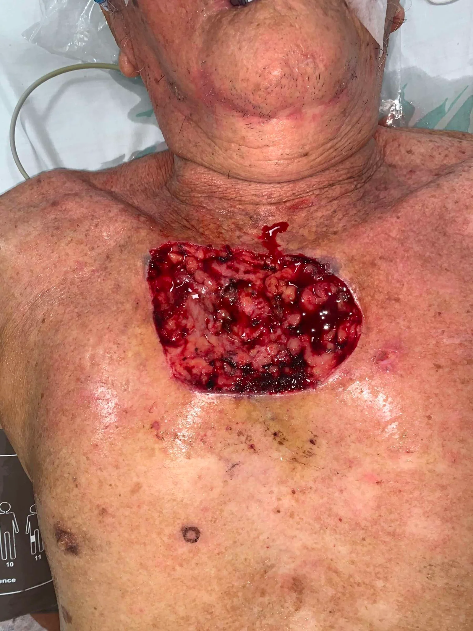
A male patient in his sixth decade of life was referred with an anterior chest wall defect measuring approximately 8 × 12 cm in diameter, resulting from resection of a squamous cell carcinoma via Mohs micrographic surgery. A propeller flap was designed, two perforators were identified intraoperatively, microsurgical dissection was performed, and flap rotation was executed.
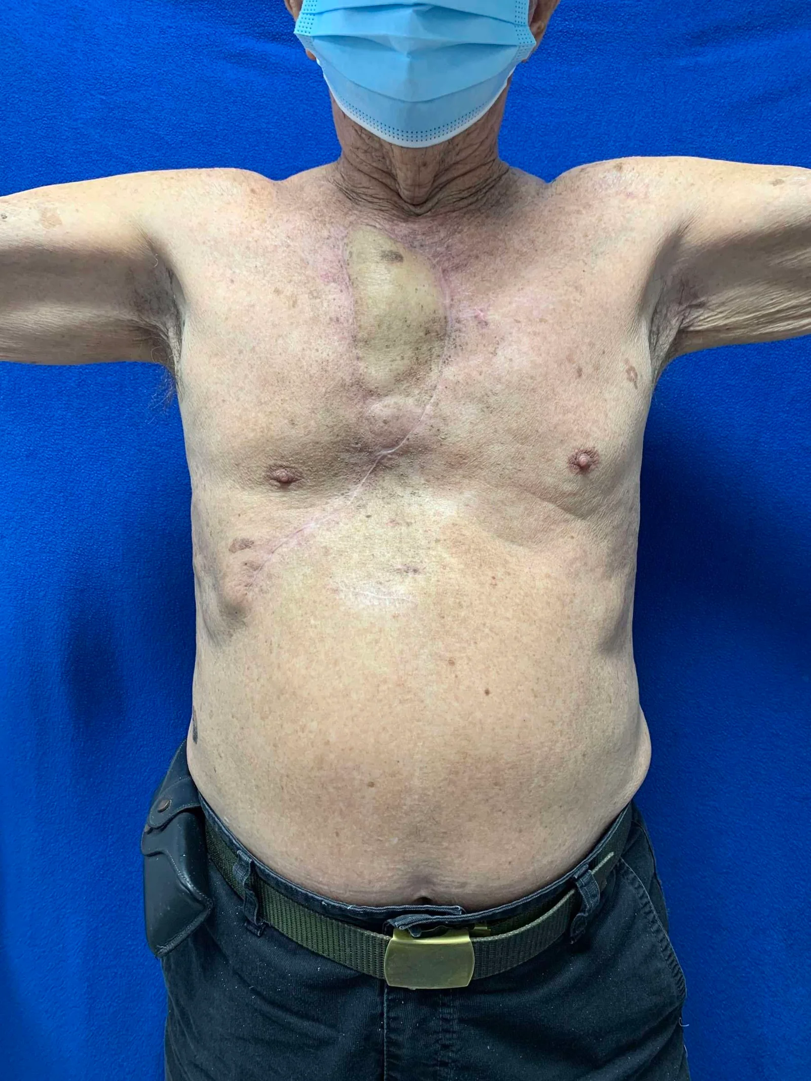
Before and After
Patient examination
chest wall defect measuring approximately 8 × 12 cm in diameter with imminent
risk of osseous exposure.
Pre-operative considerations
reconstructive goals were defined as follows:
-
Provision of durable and stable
soft tissue coverage -
Avoidance of additional
donor-site morbidity -
Prevention of secondary
deformity at the donor site

Flap Dissection
design was planned in this configuration for two reasons: 1) alignment with the
anatomical axis of the internal mammary artery perforators; and 2) donor site
closure oriented to prevent additional contour deformity.

Perforator Selection
were selected based on: 1) palpable Doppler-confirmed pulse; and 2) proximity
to the pivot point to allow adequate arc of rotation.

Isolated Perforators
meticulous dissection of the selected perforators, complete flap rotation was
achieved without vascular kinking or compromise of pedicle perfusion.

Reconstruction and Donor Site Closure
was inset into the defect, adequate perfusion was confirmed clinically, and
primary closure of the donor site was performed.
Pearls
microvascular dissection of the perforator vessels is of paramount importance
in this type of flap, particularly when the design relies on a dual-pedicle
configuration.
Pitfalls
intraoperative assessment must always be performed to ensure the absence of
perforator kinking, which may critically impair vascular inflow and compromise
flap viability.
Post-operative plan
was admitted for approximately one week of inpatient monitoring, during which
flap perfusion was assessed serially and potential complications threatening
flap viability — including hematoma formation — were actively excluded. The
patient was instructed to maintain the supine decubitus position during sleep,
and early mobilization of the upper extremities was initiated. Once wound
healing was confirmed, initiation of adjuvant radiotherapy was to proceed as
indicated per the multidisciplinary oncological treatment plan.
References
Internal mammary artery perforator (IMAP) flaps Ellis, Marco F. Operative Techniques in Otolaryngology-Head and Neck Surgery, Volume 30, Issue 2, 96 – 100
Takeuchi M, Sakurai H. Internal mammary artery perforator flap for reconstruction of the chest wall. J Plast Surg Hand Surg. 2013 Sep;47(4):328-30. doi: 10.3109/2000656X.2012.718893. Epub 2013 Jul 15. PMID: 23848424.
Rüegg EM, Lantieri L, Marchac A. Dual perforator propeller internal mammary artery perforator (IMAP) flap for soft-tissue defect of the contralateral clavicular area. J Plast Reconstr Aesthet Surg. 2012 Oct;65(10):1414-7. doi: 10.1016/j.bjps.2012.03.009. Epub 2012 Apr 13. PMID: 22503312.